. 2020 Dec;37(6):637-648.
doi: 10.1007/s10585-020-10055-x. Epub 2020 Sep 12.

Nintedanib and a bi-specific anti-VEGF/Ang2 nanobody selectively prevent brain metastases of lung adenocarcinoma cells

Affiliations

Nintedanib and a bi-specific anti-VEGF/Ang2 nanobody selectively prevent brain metastases of lung adenocarcinoma cells

Bogdana Kovalchuk et al. Clin Exp Metastasis. 2020 Dec.

Abstract

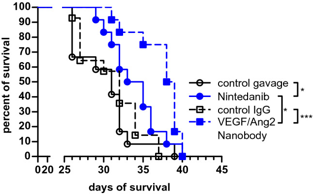
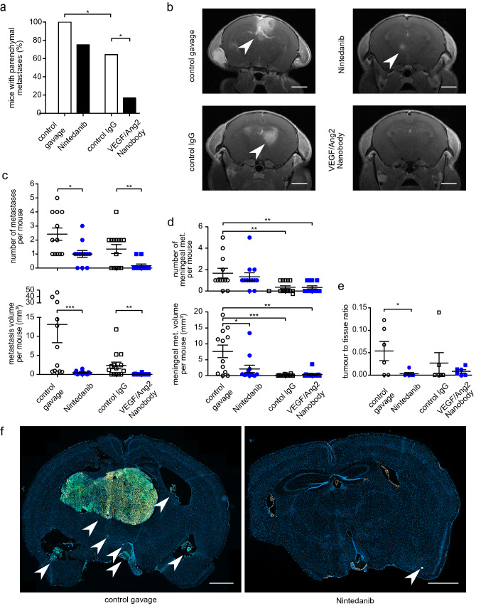
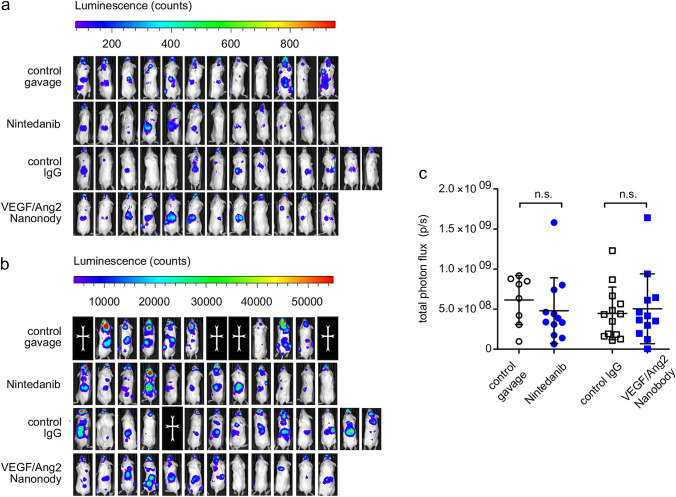
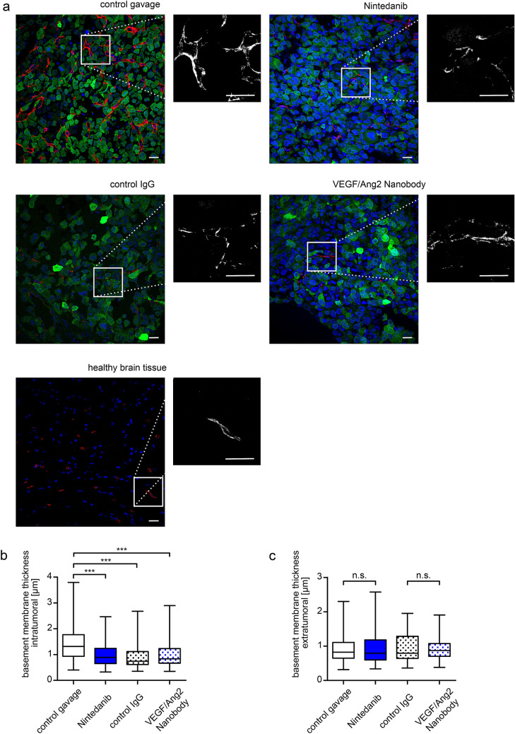
Brain metastases (BM) are an ever-increasing challenge in oncology, threatening quality of life and survival of many cancer patients. The majority of BM originate from lung adenocarcinoma, and stage III patients have a risk of 40-50% to develop BM in the first years of disease onset. As therapeutic options are limited, prevention of their occurrence is an attractive concept. Here we investigated whether Nintedanib (BIBF 1120), a tyrosine kinase inhibitor (TKI) targeting the VEGF pathway approved for lung adenocarcinoma, and the dual anti-VEGF-A/Ang2 nanobody BI836880 have the potential to prevent BM formation. A mouse model of brain metastasis from lung adenocarcinoma was used in which tumor cells were injected intracardially. Metastases formation occurred inside and outside of the brain and was followed by MRI, IVIS, and immunohistochemistry. BM were reduced in volume and number by both Nintedanib and the dual anti-VEGF-A/Ang2 nanobody, which translated into improved survival. Both compounds were able to normalize cerebral blood vessels at the site of brain metastatic lesions. Extracranial metastases, however, were not reduced, and meningeal metastases only partially. Interestingly, unspecific control IgG also lead to brain vessel normalization and reduction of brain and meningeal metastases. This data indicates a brain-specific group effect of antiangiogenic compounds with respect to metastasis prevention, most likely by preventing an early angiogenic switch. Thus, Nintedanib and BI836880 are promising candidates for future BM preventive study concepts in lung adenocarcinoma patients.

Keywords: Ang-2; Anti-angiogenic drugs; Brain neoplasms; Cancer prevention; Lung adenocarcinoma; Therapeutic IgG; VEGF-A; Xenograft metastasis model.


Conflict of interest statement

FW received a research grant from Boehringer for conducting this work. FW also received research grants from Roche, Genentech, GSK, and Divide & Conquer Ltd. ASB, MOB, MAK, MP and BK have no conflict of interest to disclose.

Figures

Fig. 1
Fig. 1
Nintedanib and VEGF/Ang2 nanobody prolong animal survival. Kaplan–Meier survival curves of the four treatment groups receiving control gavage, Nintedanib, control IgG, or VEGF/Ang2 nanobody. Median survival: 32 days; n = 12 mice per group, except of control IgG group n = 14. *p < 0.05 and ***p < 0.001 Gehan–Breslow–Wilcoxon test
Fig. 2
Fig. 2
Nintedanib and anti-VEGF/Ang2 nanobody prevent brain metastases formation. 9.4 T MRI after Gadolinium contrast administration was performed on day 26 after intracardial tumor cell injection. a Bar chart illustrating the reduced percentage of mice with metastases, detectable in cMRI. b Representative T1-w cMRI images depicting larger intracranial metastases in the control groups, indicated by arrowheads. scale bar = 2 mm. c Scatter plots showing the reduction of number and volume of cranial metastases in cMRI. d Scatter plots depicting the number and volume of meningeal metastases in cMRI e Quantification demonstrating a higher histological tumor–tissue ratio in control mice at their time of death. f Representative histological slices with higher number and size of PC14-PE6 pGF1 Br4 metastases in the control group. Fluorescent staining: DAPI (blue) = nucleus, GFP (green) = PC14-PE6 tumor cells, Alexa Flour 546 (orange) = collagen-IV positive vascular basement membrane. Arrows indicate GFP-positive metastatic lesions. Scale bar = 1000 µm. Mean values with standard errors of the mean are shown. *p < 0.05, **p < 0.01, ***p < 0.001, Mann–Whitney-U test. (Color figure online)
Fig. 3
Fig. 3
Extracranial systemic metastases are not reduced by antiangiogenic treatments. a, b IVIS luminescence images on day 14 (a) and day 28 (b) after tumor cell heart injection. Photon count visualized by heat map. c Scatter plot quantifying the extracranial photon flux on day 28 shows no difference between the treatment groups
Fig. 4
Fig. 4
Effects of drug treatment on tumor and brain microvessels. a Collagen IV basement membrane staining. Confocal images of representative intratumoral regions of mice from the four treatment groups at time of death. One extratumoral image of healthy brain tissue is also displayed. Nuclear DAPI staining is shown in blue, collagen IV staining in red (Alexa Flour 546) and GFP-expressing PC14-PE6 tumor cells in green. Vascular basement membrane signal (collagen IV) is additionally shown in single-channel images. Scale bar = 20 µm, magnification 40x. Brightness adjustments were applied equally to all images. b Scatter plot of collagen IV immunostaining inside of metastatic brain lesions. c Scatter plot of collagen IV staining of healthy brain tissue. 3 regions per mouse in 3 mice per group were analyzed. ***p < 0.001, Mann–Whitney-U test. Whiskers indicate minimum and maximum values. (Color figure online)

References

    1. Nayak L, Lee EQ, Wen PY. Epidemiology of brain metastases. Curr Oncol Rep. 2012;14(1):48–54. doi: 10.1007/s11912-011-0203-y. - DOI - PubMed
    1. Bachmann C, Schmidt S, Staebler A, Fehm T, Fend F, Schittenhelm J, Wallwiener D, Grischke E. CNS metastases in breast cancer patients: prognostic implications of tumor subtype. Med Oncol. 2014;32(1):400. doi: 10.1007/s12032-014-0400-2. - DOI - PubMed
    1. Valiente M, Ahluwalia MS, Boire A, Brastianos PK, Goldberg SB, Lee EQ, Le Rhun E, Preusser M, Winkler F, Soffietti R. The evolving landscape of brain metastasis. Trends Cancer. 2018;4(3):176–196. doi: 10.1016/j.trecan.2018.01.003. - DOI - PMC - PubMed
    1. Mamon HJ, Yeap BY, Janne PA, Reblando J, Shrager S, Jaklitsch MT, Mentzer S, Lukanich JM, Sugarbaker DJ, Baldini EH, Berman S, Skarin A, Bueno R. High risk of brain metastases in surgically staged IIIA non-small-cell lung cancer patients treated with surgery, chemotherapy, and radiation. J Clin Oncol. 2005;23(7):1530–1537. doi: 10.1200/JCO.2005.04.123. - DOI - PubMed
    1. Peters S, Camidge DR, Shaw AT, Gadgeel S, Ahn JS, Kim DW, Ou SI, Perol M, Dziadziuszko R, Rosell R, Zeaiter A, Mitry E, Golding S, Balas B, Noe J, Morcos PN, Mok T, Investigators AT. Alectinib versus crizotinib in untreated ALK-positive non-small-cell lung cancer. N Engl J Med. 2017;377(9):829–838. doi: 10.1056/NEJMoa1704795. - DOI - PubMed

Publication types

MeSH terms