. 2020 Nov 1:260:118400.
doi: 10.1016/j.lfs.2020.118400. Epub 2020 Sep 9.

Parameters predicting COVID-19-induced myocardial injury and mortality

Affiliations

Parameters predicting COVID-19-induced myocardial injury and mortality

G D Duerr et al. Life Sci. .

Abstract

Clinical manifestations of COVID-19 affect many organs, including the heart. Cardiovascular disease is a dominant comorbidity and prognostic factors predicting risk for critical courses are highly needed. Moreover, immunomechanisms underlying COVID-induced myocardial damage are poorly understood.

Objective: To elucidate prognostic markers to identify patients at risk.

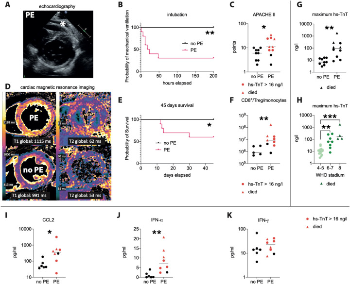
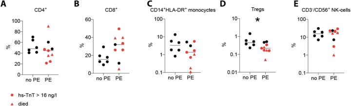
Results: Only patients with pericardial effusion (PE) developed a severe disease course, and those who died could be identified by a high CD8/Treg/monocyte ratio. Ten out of 19 COVID-19 patients presented with PE, 7 (78%) of these had elevated APACHE-II mortality risk-score, requiring mechanical ventilation. At admission, PE patients showed signs of systemic and cardiac inflammation in NMR and impaired cardiac function as detected by transthoracic echocardiography (TTE), whereas parameters of myocardial injury e.g. high sensitive troponin-t (hs-TnT) were not yet increased. During the course of disease, hs-TnT rose in 8 of the PE-patients above 16 ng/l, 7 had to undergo ventilatory therapy and 4 of them died. FACS at admission showed in PE patients elevated frequencies of CD3+CD8+ T cells among all CD3+ T-cells, and lower frequencies of Tregs and CD14+HLA-DR+-monocytes. A high CD8/Treg/monocyte ratio predicted a severe disease course in PE patients, and was associated with high serum levels of antiviral cytokines. By contrast, patients without PE and PE patients with a low CD8/Treg/monocyte ratio neither had to be intubated, nor died.

Conclusions: PE predicts cardiac injury in COVID-19 patients. Therefore, TTE should be performed at admission. Immunological parameters for dysfunctional antiviral immunity, such as the CD8/Treg/monocyte ratio used here, supports risk assessment by predicting poor prognosis.

Keywords: CD8; COVID-19; Monocyte; Pericardial effusion; SARS-CoV-2; Treg.


Conflict of interest statement

The authors report no conflicts of interest.

Figures

Fig. 1
Fig. 1
PE, hs-TnT and the CD8/Treg/monocyte quotient predict heart injury and clinical course of COVID-19 patients. (A): representative transthoracic echocardiography (TTE) image, asterisk indicates pericardial effusion. (B): necessity for intubation, (C) APACHE II-score. (D): Representative cardiac magnetic resonance images from a patient with (upper row) and without PE (lower row), showing a diffuse inflammatory pattern with increased markers of edema (T1 and T2 relaxation times) only in PE patients. (E) Survival of COVID-19 patients with or without pericardial effusion (PE) at admission, showing that a PE predicts poorer prognosis. (F): ratio between CD8+ T cells to Tregs and CD14+ HLA-DR+ monocytes, as a parameter for disease severity, which was higher in patients with PE, especially in those who subsequently developed a hs-TnT rise (red symbols) and those who died (red triangles). (G): hs-TnT correlates with PE and with disease severity indicated by (H) the new WHO COVID-19 classification. (I–K): Serum concentrations of the antiviral cytokines CCL2 (I), IFN-α (J) and IFN-γ (K) in patients exhibiting a pericardial effusion (PE) or not (no PE). Subsequent hs-TnT rise (red symbols), deceased (red triangle). Data is presented as scatter plot with median (line). p < 0.05 was considered statistically significant (*). **, p < 0.01; ***, p < 0.001. (For interpretation of the references to color in this figure legend, the reader is referred to the web version of this article.)
Fig. 2
Fig. 2
Immune cell blood frequencies in COVID-19 patients. Percentages of CD4+ of CD3+ T cells (A), CD8+ of CD3+ T cells (B), CD14+ HLA-DR+ monocytes (C), Tregs (D) and NK cells (E) upon admission in patients exhibiting a pericardial effusion (PE) versus patients without PE (no PE).

References

    1. Shi S., Qin M., Cai Y., Liu T., Shen B., Yang F., Cao S., Liu X., Xiang Y., Zhao Q., Huang H., Yang B., Huang C. Characteristics and clinical significance of myocardial injury in patients with severe coronavirus disease 2019. Eur. Heart J. 2020;41:2070–2079. - PMC - PubMed
    1. Doyen D., Moceri P., Ducreux D., Dellamonica J. Myocarditis in a patient with COVID-19: a cause of raised troponin and ECG changes. Lancet. 2020;395:1516. - PMC - PubMed
    1. Fox S.E., Li G., Akmatbekov A., Harbert J.L., Lameira F.S., Brown J.Q., Vander Heide R.S. Unexpected features of cardiac pathology in COVID-19 infection. Circulation. 2020 doi: 10.1161/CIRCULATIONAHA.120.049465. - DOI - PubMed
    1. Babapoor-Farrokhran S., Gill D., Walker J., Rasekhi R.T., Bozorgnia B., Amanullah A. Myocardial injury and COVID-19: possible mechanisms. Life Sci. 2020;253 - PMC - PubMed
    1. Libby P. The heart in COVID19: primary target or secondary bystander? JACC Basic Transl. Sci. 2020;5(5):537–542. doi: 10.1016/j.jacbts.2020.04.001. eCollection 2020 May. - DOI - PMC - PubMed

MeSH terms