. 2020 Dec:42:101078.
doi: 10.1016/j.molmet.2020.101078. Epub 2020 Sep 9.

Wnt/β-catenin signaling regulates adipose tissue lipogenesis and adipocyte-specific loss is rigorously defended by neighboring stromal-vascular cells

Affiliations

Wnt/β-catenin signaling regulates adipose tissue lipogenesis and adipocyte-specific loss is rigorously defended by neighboring stromal-vascular cells

Devika P Bagchi et al. Mol Metab. 2020 Dec.

Abstract

Objective: Canonical Wnt/β-catenin signaling is a well-studied endogenous regulator of mesenchymal cell fate determination, promoting osteoblastogenesis and inhibiting adipogenesis. However, emerging genetic evidence in humans links a number of Wnt pathway members to body fat distribution, obesity, and metabolic dysfunction, suggesting that this pathway also functions in adipocytes. Recent studies in mice have uncovered compelling evidence that the Wnt signaling pathway plays important roles in adipocyte metabolism, particularly under obesogenic conditions. However, complexities in Wnt signaling and differences in experimental models and approaches have thus far limited our understanding of its specific roles in this context.

Methods: To investigate roles of the canonical Wnt pathway in the regulation of adipocyte metabolism, we generated adipocyte-specific β-catenin (β-cat) knockout mouse and cultured cell models. We used RNA sequencing, ChIP sequencing, and molecular approaches to assess expression of Wnt targets and lipogenic genes. We then used functional assays to evaluate effects of β-catenin deficiency on adipocyte metabolism, including lipid and carbohydrate handling. In mice maintained on normal chow and high-fat diets, we assessed the cellular and functional consequences of adipocyte-specific β-catenin deletion on adipose tissues and systemic metabolism.

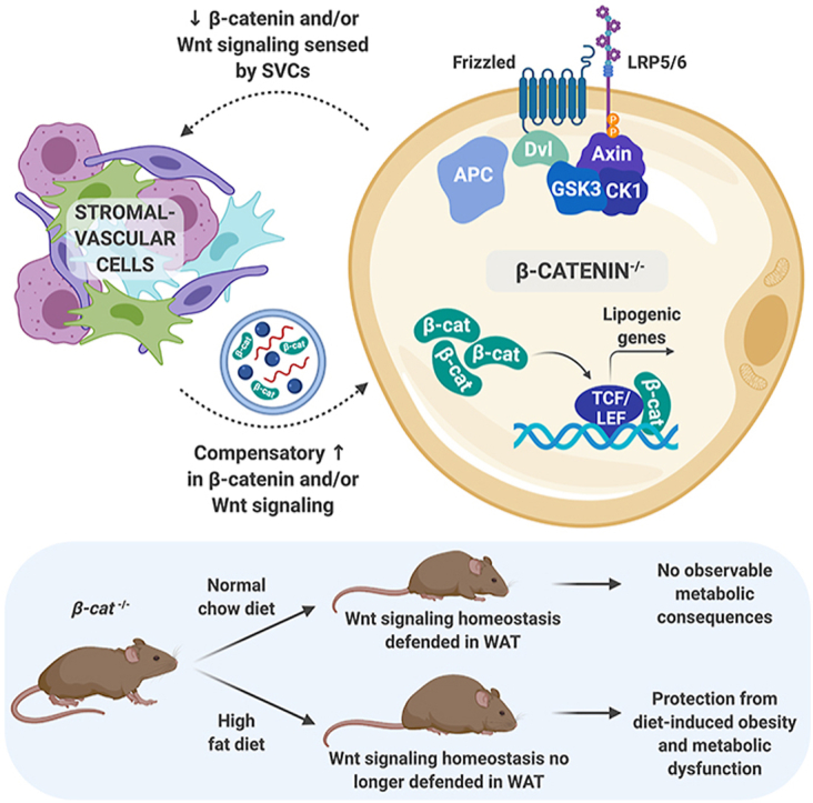
Results: We report that in adipocytes, the canonical Wnt/β-catenin pathway regulates de novo lipogenesis (DNL) and fatty acid monounsaturation. Further, β-catenin mediates effects of Wnt signaling on lipid metabolism in part by transcriptional regulation of Mlxipl and Srebf1. Intriguingly, adipocyte-specific loss of β-catenin is sensed and defended by CD45-/CD31- stromal cells to maintain tissue-wide Wnt signaling homeostasis in chow-fed mice. With long-term high-fat diet, this compensatory mechanism is overridden, revealing that β-catenin deletion promotes resistance to diet-induced obesity and adipocyte hypertrophy and subsequent protection from metabolic dysfunction.

Conclusions: Taken together, our studies demonstrate that Wnt signaling in adipocytes is required for lipogenic gene expression, de novo lipogenesis, and lipid desaturation. In addition, adipose tissues rigorously defend Wnt signaling homeostasis under standard nutritional conditions, such that stromal-vascular cells sense and compensate for adipocyte-specific loss. These findings underscore the critical importance of this pathway in adipocyte lipid metabolism and adipose tissue function.

Keywords: Adipocyte; Adipose tissue; Lipogenesis; Metabolism; Wnt signaling; β-catenin.


Figures

Image 1
Graphical abstract
Figure 1
Figure 1
β-catenin is expressed in cultured and primary adipocytes and up-regulated by diet-induced obesity. (A-B) Mesenchymal stem cells (MSC) isolated from C57BL/6J mice were cultured under standard conditions and induced to differentiate. Ctnnb1 gene (n = 6) and protein (n = 2) expression at indicated days of adipogenesis. (C) Ctnnb1 gene expression in stromal-vascular (SVF) and adipocyte (Ads) fractions isolated from epididymal (eWAT) and inguinal (iWAT) white adipose tissues (WAT) of C57BL/6J mice (males; n = 5). (D) Expression of Ctnnb1 in eWAT and iWAT of mice fed a normal chow diet (NCD) or high-fat diet (HFD) for 10 weeks. (E) Ctnnb1 expression in SVF and Ads of eWAT and iWAT isolated from NCD- and HFD-fed mice (males; n = 6). (F) Ctnnb1 allele structure and genetic recombination in β-catfl/fl and β-cat−/− adipocytes using a 3-primer PCR system (n = 3). (G-H) Ctnnb1 RNA (n = 6) and protein (n = 3) expression in adipocytes following adenoviral GFP or Cre infection. (I) Representative brightfield and Oil Red O images, and (J) triacylglycerol (TAG) accumulation in β-catfl/fl and β-cat−/− adipocytes (n = 6). (K) Expression of Ctnnb1 and downstream Wnt target genes in β-catfl/fl and β-cat−/− adipocytes treated with vehicle or 3 μM CHIR99021 for 4 h (n = 6). RNA expression normalized to PPIA. Data presented as mean ± S.D. ∗ indicates significance at p < 0.05.
Figure 2
Figure 2
β-catenin regulates metabolic pathways in adipocytes and exclusively mediates effects of canonical Wnt3a signaling. RNA-seq analyses were performed on β-catfl/fl and β-cat−/− adipocytes under basal conditions or after 4 h of treatment with recombinant Wnt3a (20 ng/ml; n = 4 per group). (A) Heat maps of differential gene expression changes in β-catfl/fl and β-cat−/− adipocytes under basal conditions (left panel) and β-catfl/fl cells treated with vehicle or Wnt3a (right panel). (B) Gene Set Enrichment Analyses (GSEA) of genes expressed in β-catfl/fl and β-cat−/− adipocytes under basal conditions (top panel) and β-catfl/fl cells treated with vehicle or Wnt3a (bottom panel). (C-D) MA plots of gene expression changes following Wnt3a treatment of β-catfl/fl or β-cat−/− adipocytes. (E) Venn diagram depicting meta-analysis of gene expression changes in β-catfl/fl or β-cat−/− adipocytes treated with vehicle or Wnt3a for 4 h. (F) Expression of Ctnnb1 and downstream Wnt target genes in β-catfl/fl and β-cat−/− adipocytes treated with vehicle or 20 ng/ml recombinant Wnt3a for 4, 12, or 24 h (n = 6). RNA expression normalized to PPIA. Data presented as mean ± S.D. ∗ indicates significance at p < 0.05.
Figure 3
Figure 3
β-catenin-dependent Wnt signaling regulates lipogenesis and fatty acid desaturation in adipocytes. (A-B) Heat map and MA plot showing differentially expressed genes related to fatty acid, cholesterol, and bile acid metabolism in cultured β-catfl/fl and β-cat−/− adipocytes (n = 4). (C-D) Lipogenic gene (n = 6) and protein (n = 3) expression in β-catfl/fl and β-cat−/− adipocytes. (E) Proportion of total saturated vs unsaturated fatty acids in lipids extracted from β-catfl/fl and β-cat−/− adipocytes (n = 3). (F) Relative proportions of myristic (C14:0) and palmitic (C16:0) vs myristoleic (C14:1, n-5) and palmitoleic (C16:1, n-7) acids (n = 3). (G) De novo lipogenesis (DNL) was evaluated in cultured β-catfl/fl and β-cat−/− adipocytes using [14C]-acetate for 2, 4, and 8 h. Incorporation of [14C]-radiolabel into TAG fractions extracted from β-catfl/fl and β-cat−/− adipocytes was quantified by scintillation counting (n = 6). (H–I) Gene (n = 6) and protein (n = 3) expression of indicated transcription factors in β-catfl/fl and β-cat−/− adipocytes. (J) Protein expression in β-catfl/fl and β-cat−/− adipocytes treated for 72 h with adenovirus expressing GFP, ChREBP, or SREBP1c (1 × 105 viral particles/ml). (K) Integrative Genomics Viewer capture showing Tcf7l2 peaks (indicating binding occupancy) in regions ± 3 kb from transcription start sites (black arrows) of the indicated genes in cultured Tcf7l2fl/fl and Tcf7l2−/− adipocytes. RNA expression normalized to PPIA. Data presented as mean ± S.D. ∗ indicates significance at p < 0.05.
Figure 4
Figure 4
Adipocyte-specific β-catenin deletion does not influence global metabolism on a normal chow diet. (A) Genetic recombination in tissues isolated from β-cat−/− mice. (B) Growth curves of 28-week-old β-catfl/fl and β-cat−/− mice. (C) Body composition of 16-week-old β-catfl/fl and β-cat−/− mice on NCD. (D) Glucose tolerance test in 16-week-old β-catfl/fl and β-cat−/− mice. (E) Insulin tolerance test in 19-week-old mice. (F) Blood glucose concentrations in random-fed and 16 h fasted mice. Serum concentrations of (G) random-fed and fasted insulin and (H) adiponectin levels in 28-week-old mice. (I) Basal and stimulated lipolysis in 22-week-old mice (iso, isoproterenol: 10 mg/kg body weight). (J) Serum TAG in 28-week-old mice. (K) Tissue weights at time of sacrifice. (L) Representative histological images of H&E-stained tissues from β-catfl/fl and β-cat−/− mice fed NCD for 28 weeks; 200x magnification; scale bar, 100 μm. Data in B-L from male mice, n = 8 per group. Data presented as mean ± S.D. ∗ indicates significance at p < 0.05.
Figure 5
Figure 5
β-catenin is up-regulated in the stromal-vascular fraction of adipose tissues from knockout mice. (A) Genetic recombination in tissues isolated from β-catfl/fl and β-cat−/− mice (n = 3). (B–C) Ctnnb1 mRNA (n = 3) and protein (n = 6) expression in eWAT and iWAT of β-catfl/fl and β-cat−/− mice. (D) Genomic recombination of β-catenin in adipocytes and SVF isolated from eWAT and iWAT of β-catfl/fl and β-cat−/− mice. (E) Ctnnb1 mRNA expression in isolated eWAT adipocytes and SVF of β-catfl/fl and β-cat−/− mice (n = 6). (F) β-catenin protein expression in isolated eWAT adipocytes and SVF of β-catfl/fl and β-cat−/− mice; adiponectin and laminin shown as protein loading controls. (G-H) Wnt target gene expression in adipocytes and SVF isolated from eWAT of β-catfl/fl and β-cat−/− mice (n = 8). (I) Representative plots showing flow cytometry analysis of SVF isolated from β-catfl/fl and β-cat−/− mice (3 mice per sample; n = 3 samples). (J) Quantification of SVF cell proportions evaluated by flow cytometry analysis (3 mice per sample; n = 3 samples). (K) Ctnnb1 mRNA expression normalized to PPIA in cellular fractions isolated by FACS analysis (3 mice per sample; n = 3 samples). Data presented as mean ± S.D. ∗ indicates significance at p < 0.05.
Figure 6
Figure 6
β-cat−/−mice are protected from diet-induced obesity and metabolic dysfunction. (A) Growth curves over time of 32-week-old β-catfl/fl and β-cat−/− mice fed 60% HFD for 24 weeks. (B) Body composition analysis of 28-week-old mice. (C-D) Glucose tolerance test and area under the curve analysis in 28-week-old mice. (E) Blood glucose concentrations in random-fed and 16 h fasted mice. (F) Insulin tolerance test in 30-week-old mice. (G) Serum insulin concentrations in random-fed mice or 16 h fasted mice at indicated times after intraperitoneal glucose injection (1 mg/kg body weight). Serum (H) TAG, (I) total cholesterol, and (J) adiponectin in 32-week-old mice. (K) Tissue weights at time of sacrifice. (L) Representative histological images of H&E-stained tissues from β-catfl/fl and β-cat−/− mice fed HFD for 24 weeks; 200x magnification; scale bar, 100 μm. (M) Quantification of liver TAG in 32-week-old mice. Data shown from male mice; β-catfl/fl: n = 5, β-cat−/−: n = 11. Data presented as mean ± S.D. ∗ indicates significance at p < 0.05.
Figure 7
Figure 7
Diet-induced obesity overcomes compensatory up-regulation of Wnt/β-catenin signaling in the SVF of knockout mice. (A) Ctnnb1 mRNA expression in eWAT and iWAT of β-catfl/fl and β-cat−/− mice fed HFD for 28 weeks (n = 5). (B) Ctnnb1 mRNA expression in isolated eWAT adipocytes and SVF of HFD-fed β-catfl/fl and β-cat−/− mice (n = 5). (C) β-catenin protein expression in isolated eWAT adipocytes and SVF of β-catfl/fl and β-cat−/− mice fed HFD; adiponectin and laminin shown as controls. (D-E) Wnt target gene expression in SVF and adipocytes isolated from eWAT of obese β-catfl/fl and β-cat−/− mice (n = 5). (F) Lipogenic gene expression in eWAT adipocytes isolated from HFD-fed β-catfl/fl and β-cat−/− mice (n = 8). (G) Expression of immune, endothelial, and stromal cell markers in SVF isolated from eWAT of obese β-catfl/fl and β-cat−/− mice (n = 8). (H–I) Expression of inflammatory markers in whole eWAT and iWAT of β-catfl/fl and β-cat−/− mice fed HFD (n = 8). RNA expression normalized to PPIA. Data presented as mean ± S.D. ∗ indicates significance at p < 0.05.

Similar articles

Cited by

References

    1. Cadigan K.M., Nusse R. Wnt signaling: a common theme in animal development. Genes & Development. 1997;11(24):3286–3305. - PubMed
    1. Clevers H. Wnt/beta-catenin signaling in development and disease. Cell. 2006;127(3):469–480. - PubMed
    1. Logan C.Y., Nusse R. The Wnt signaling pathway in development and disease. Annual Review of Cell and Developmental Biology. 2004;20:781–810. - PubMed
    1. MacDonald B.T., Tamai K., He X. Wnt/beta-catenin signaling: components, mechanisms, and diseases. Developmental Cell. 2009;17(1):9–26. - PMC - PubMed
    1. Valenta T., Hausmann G., Basler K. The many faces and functions of beta-catenin. The EMBO Journal. 2012;31(12):2714–2736. - PMC - PubMed

Publication types

MeSH terms

Substances