Review
. 2020 Sep:124:103960.
doi: 10.1016/j.compbiomed.2020.103960. Epub 2020 Aug 14.

COVID-19 pathways for brain and heart injury in comorbidity patients: A role of medical imaging and artificial intelligence-based COVID severity classification: A review

Affiliations
Review

COVID-19 pathways for brain and heart injury in comorbidity patients: A role of medical imaging and artificial intelligence-based COVID severity classification: A review

Jasjit S Suri et al. Comput Biol Med. 2020 Sep.

Abstract

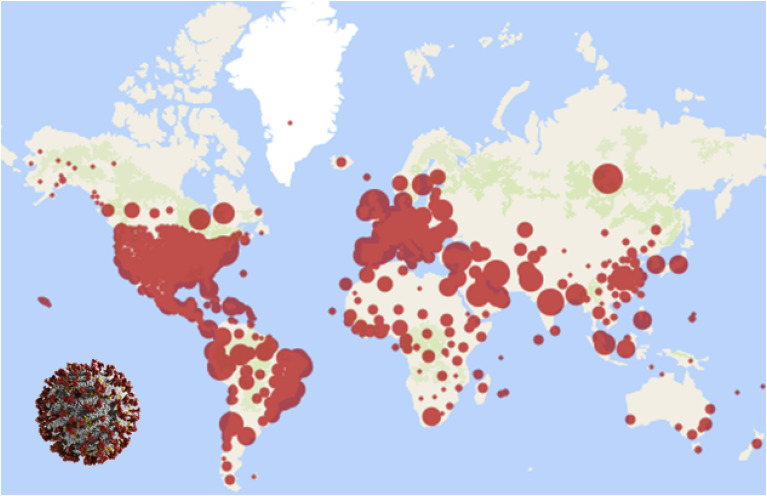
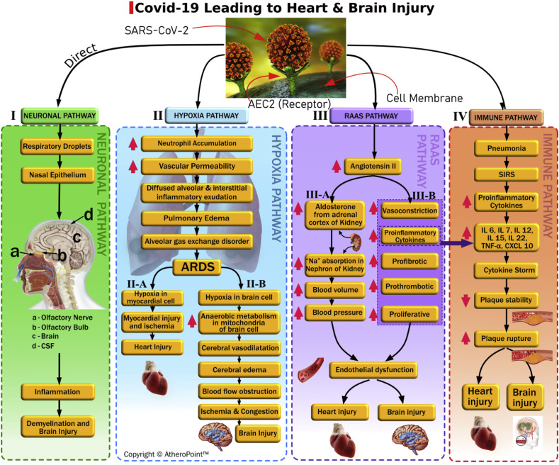
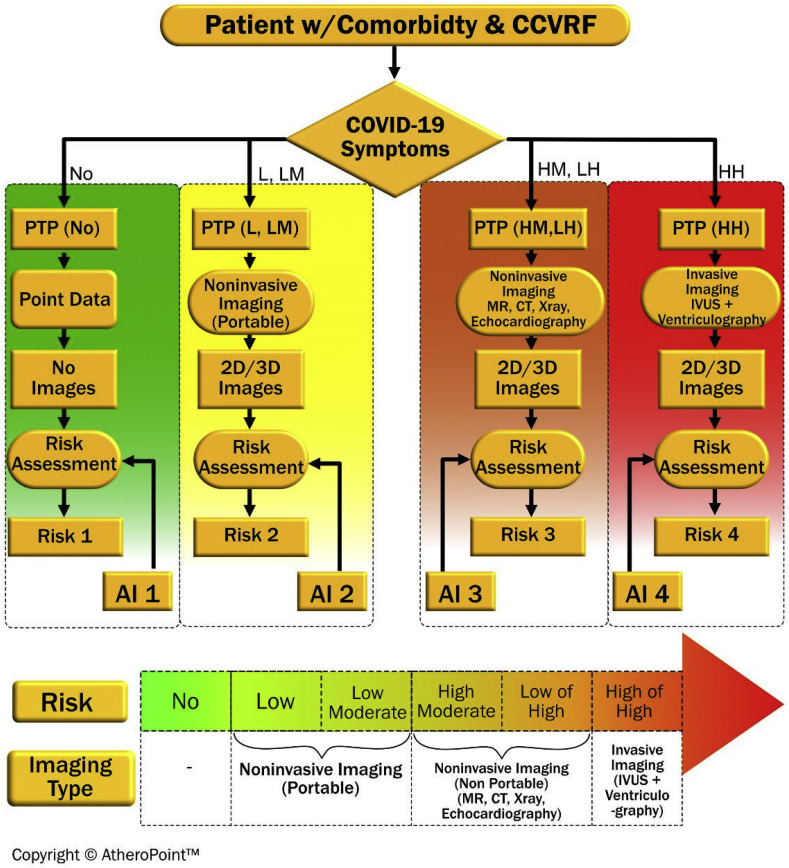
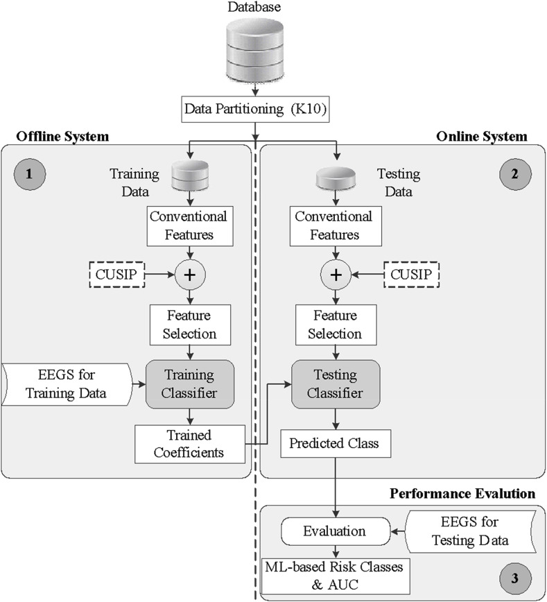
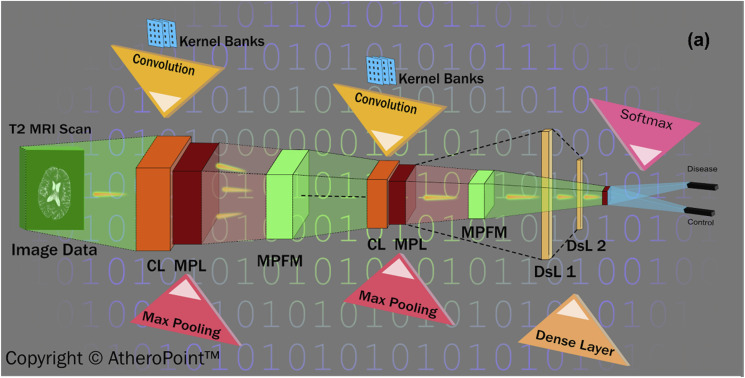
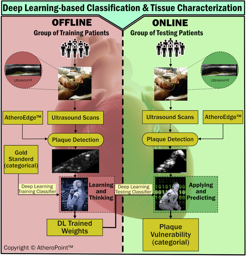
Artificial intelligence (AI) has penetrated the field of medicine, particularly the field of radiology. Since its emergence, the highly virulent coronavirus disease 2019 (COVID-19) has infected over 10 million people, leading to over 500,000 deaths as of July 1st, 2020. Since the outbreak began, almost 28,000 articles about COVID-19 have been published (https://pubmed.ncbi.nlm.nih.gov); however, few have explored the role of imaging and artificial intelligence in COVID-19 patients-specifically, those with comorbidities. This paper begins by presenting the four pathways that can lead to heart and brain injuries following a COVID-19 infection. Our survey also offers insights into the role that imaging can play in the treatment of comorbid patients, based on probabilities derived from COVID-19 symptom statistics. Such symptoms include myocardial injury, hypoxia, plaque rupture, arrhythmias, venous thromboembolism, coronary thrombosis, encephalitis, ischemia, inflammation, and lung injury. At its core, this study considers the role of image-based AI, which can be used to characterize the tissues of a COVID-19 patient and classify the severity of their infection. Image-based AI is more important than ever as the pandemic surges and countries worldwide grapple with limited medical resources for detection and diagnosis.

Keywords: Artificial intelligence; Brain; COVID-19; Comorbidity; Heart; Imaging; Lung; Pathophysiology; Risk assessment.


Conflict of interest statement

The authors declare that they have no known competing financial interests or personal relationships that could have appeared to influence the work reported in this paper.

Figures

Fig. 1
Fig. 1
World map showing COVID-19 spread over 213 countries (courtesy: John Hopkins University).
Fig. 2
Fig. 2
(a) Association of SARS-CoV-2, with other comorbidities, and (b) comparison of the mortality rate of diabetic and non-diabetic COVID-19 patients (reproduced with permission [11]).
Fig. 3
Fig. 3
We have shown in four pathways how COVID-19 can cause Brain and heart injury. Brain image in pathway I: http://debuglies.com/2020/01/23/olfactory-disturbances-have-implications-in-mental-and-emotional-well-being-health/(Courtesy of Debug Lies).
Fig. 4
Fig. 4
MRI scan of COVID-19 patient showing hemorrhage. MRI images demonstrate T2 FLAIR hyperintensity within the bilateral medial temporal lobes and thalami (A, B, E, F) with evidence of hemorrhage indicated by hypointense signal intensity on susceptibility-weighted images (C, G) and rim enhancement on postcontrast images (D, H) (reproduced with permission [108]).
Fig. 5
Fig. 5
Application of chest CT and IVUS for a COVID-19 patient suffering from myocardial infarction (a) Chest CT scan with viral pneumonia showing fibrinous, focal exudative changes. (b) When the patient complained of chest pain, the ECG report showed the ST-segments elevations in V1–V5 lead. (c, d) CAG radiology that the proximal segment of LAD was occluded. (e, f) The blood flow of LAD restoration after 2 DESs was implanted. (g) The dissection distal shown by IVUS to the stent in LAD from 7 to 12 o'clock. (h) The low echogenic shadow with scattered higher echogenic flicker, indicating a thrombus. (i) After a DES was implanted, and the stent was well expanded, the dissection could not be seen (j) The thrombus disappearance after the intervention (reproduced with permission [112]).
Fig. 6
Fig. 6
Role of AI-based risk assessment on COVID-19 patients having comorbidity.
Fig. 7
Fig. 7
Typical low-cost machine learning architecture utilizing the EEGS model.
Fig. 8
Fig. 8
A convolution neural network (courtesy of AtheroPoint™, CA, USA).
Fig. 9
Fig. 9
Proposed DL-based system for tissue characterization and classification of COVID-19 severity with patients with comorbidities (courtesy of AtheroPoint™, CA, USA).
Fig. 10
Fig. 10
a) Safety guidelines to be followed by medical staff before performing imaging (reproduced with permission [188]); (b) Images being taken through glass (reproduced with permission [189]); (c) disposable sterile sheath for covering probe. (reproduced with permission [190]).

References

    1. Yuen K.-S., Ye Z.-W., Fung S.-Y., Chan C.-P., Jin D.-Y. SARS-CoV-2 and COVID-19: the most important research questions. Cell Biosci. 2020;10(1):1–5. - PMC - PubMed
    1. Coronavirus (COVID-19) outbreak [https://www.who.int/westernpacific/emergencies/covid-19].
    1. Coronavirus [https://www.who.int/health-topics/coronavirus#tab=tab_1].
    1. Chan J.F.-W., Yuan S., Kok K.-H., To K.K.-W., Chu H., Yang J., Xing F., Liu J., Yip C.C.-Y., Poon R.W.-S. A familial cluster of pneumonia associated with the 2019 novel coronavirus indicating person-to-person transmission: a study of a family cluster. Lancet. 2020;395(10223):514–523. - PMC - PubMed
    1. Coronavirus [https://www.worldometers.info/coronavirus/].

MeSH terms