

Extra digital glomus tumor: A very rare cause of chronic abdominal wall pain
- PMID: 32919334
- PMCID: PMC7490988
- DOI: 10.1016/j.ijscr.2020.09.002
Extra digital glomus tumor: A very rare cause of chronic abdominal wall pain
Abstract
Introduction: Glomus tumors are very rare benign vascular tumors, constituting less than 2% of soft tissue tumors. These tumors originate from the glomus body. 75% of these tumors occur in hand however rarely can be found in any body part.
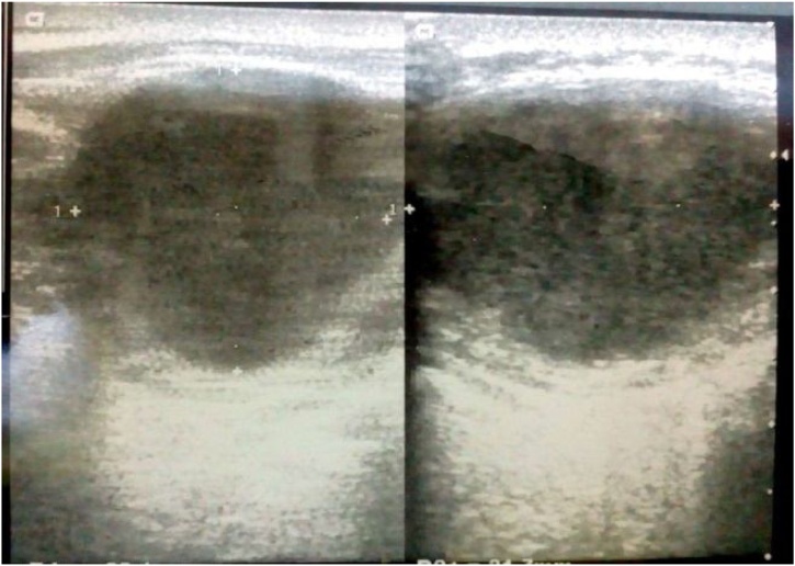
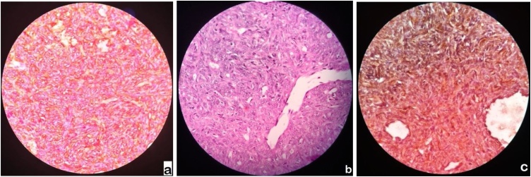
Presentation of case: We here report a case of glomus tumor who presented with abdominal pain (constant and throbbing nature) and small swelling in the left hypochondrium. Pain was mostly spontaneous without any obvious cause, aggravated by cold and palpation. Ultrasonography parietal wall showed 27 × 22 × 21 mm hypoechoic lesion in the parietal wall with increased focal vascularity. Histopathological examination confirmed the diagnosis of glomus tumor.
Discussion: These are rare benign vascular tumors arising from the glomus bodies found anywhere in body. However 75% are found in hand mostly subungual region. Glomus tumor may show unusual clinical picture such as extra digital location, large size, deep soft tissue, visceral location, multi-centric or infiltrative growth pattern. These tumor commonly presents with a diagnostic triad of spontaneous pain, hypersensitivity to drop in temperature and pressure tenderness. Clinical diagnostic tests aide in diagnosis, including Love's test, Hildreth's test, Transillumination and the cold test. The clinical differential diagnosis of glomus tumor includes Raynaud's phenomenon, neuroma, gout, infection, peripheral neuropathy and radiculopathy.
Conclusion: Extra digital glomus tumor occur in any part of the body and should be put in differential diagnosis of abdominal pain when no obvious cause of pain is found. Surgical excision is the curative treatment of choice with rare recurrence.
Keywords: Abdominal pain; Abdominal wall; Glomus tumor.
Copyright © 2020. Published by Elsevier Ltd.
Figures
Similar articles
-
Diagnosis and surgical approach in treating glomus tumor distal phalanx left middle finger: A case report.Int J Surg Case Rep. 2023 Jul;108:108426. doi: 10.1016/j.ijscr.2023.108426. Epub 2023 Jun 18. Int J Surg Case Rep. 2023. PMID: 37392587 Free PMC article.
-
Subungual glomus tumors of the hand: Treated by transungual excision.Indian J Orthop. 2015 Jul-Aug;49(4):403-7. doi: 10.4103/0019-5413.159611. Indian J Orthop. 2015. PMID: 26229160 Free PMC article.
-
Glomus tumors of the hand.Eplasty. 2008;8:e48. Epub 2008 Oct 8. Eplasty. 2008. PMID: 18997858 Free PMC article.
-
Glomus tumours of the hand: Review of literature.J Clin Orthop Trauma. 2016 Oct-Dec;7(4):286-291. doi: 10.1016/j.jcot.2016.04.006. Epub 2016 Sep 1. J Clin Orthop Trauma. 2016. PMID: 27857505 Free PMC article. Review.
-
Glomus tumor causing chronic finger pain: ischemia test is a reliable clinical sign for the diagnosis. Case report and review of literature.Rev Esp Anestesiol Reanim (Engl Ed). 2024 Apr;71(4):339-343. doi: 10.1016/j.redare.2024.02.021. Epub 2024 Feb 27. Rev Esp Anestesiol Reanim (Engl Ed). 2024. PMID: 38423461 Review.
Cited by
-
Suprascapular glomus tumor: an unusual cause of chronic shoulder pain.Case Reports Plast Surg Hand Surg. 2021 Dec 10;9(1):22-25. doi: 10.1080/23320885.2021.2014335. eCollection 2022. Case Reports Plast Surg Hand Surg. 2021. PMID: 34912941 Free PMC article.
-
Low-grade malignancy glomus tumor of the abdominal wall: a case report and literature review.J Surg Case Rep. 2023 Dec 18;2023(12):rjad680. doi: 10.1093/jscr/rjad680. eCollection 2023 Dec. J Surg Case Rep. 2023. PMID: 38115948 Free PMC article.
References
-
- Enzinger F.M., Weiss S.W. Perivascular tumors. In: Enzinger F.M., Weiss S.W., editors. Enzinger and Weiss’ Soft Tissue Tumors. 4th ed. Mosby; St Louis (MO): 2001. pp. 985–1035.
-
- Kale S.S., Rao V.K., Bentz M.L. Glomus tumor of the index finger. J. Craniofac. Surg. 2006;17:801–804. - PubMed
-
- Schiefer T.K., Parker W.L., Anakwenze O.A., Amadio P.C., Inwards C.Y., Spinner R.J. Extradigital glomus tumors: a 20-year experience. Mayo Clin. Proc. 2006;81:1337–1344. - PubMed
-
- Agha R.A., Borrelli M.R., Farwana R., Koshy K., Fowler A.J., Orgill D.P., Zhu H., Alsawadi A., Noureldin A., Rao A., Enam A. The SCARE 2018 statement: updating consensus Surgical CAse REport (SCARE) guidelines. Int. J. Surg. 2018;60(December):132–136. - PubMed
-
- Gould E.W., Manivel J.C., Albores-Saavedra J., Monteforte H. Locally infiltrative glomus tumours and glomangiosarcomas: a clinical, ultrastructural and immunohistochemical study. Cancer. 1990;65:310–318. - PubMed
Publication types
LinkOut - more resources
Full Text Sources
Research Materials
