

Network Effects of the 15q13.3 Microdeletion on the Transcriptome and Epigenome in Human-Induced Neurons
- PMID: 32919612
- PMCID: PMC9359316
- DOI: 10.1016/j.biopsych.2020.06.021
Network Effects of the 15q13.3 Microdeletion on the Transcriptome and Epigenome in Human-Induced Neurons
Erratum in
-
Errata.Biol Psychiatry. 2021 Mar 1;89(5):532. doi: 10.1016/j.biopsych.2020.09.017. Biol Psychiatry. 2021. PMID: 33541526 No abstract available.
Abstract
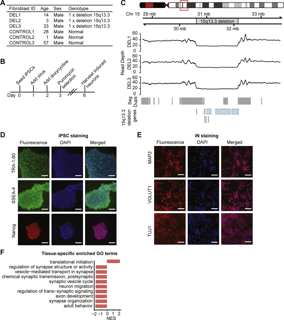
Background: The 15q13.3 microdeletion is associated with several neuropsychiatric disorders, including autism and schizophrenia. Previous association and functional studies have investigated the potential role of several genes within the deletion in neuronal dysfunction, but the molecular effects of the deletion as a whole remain largely unknown.
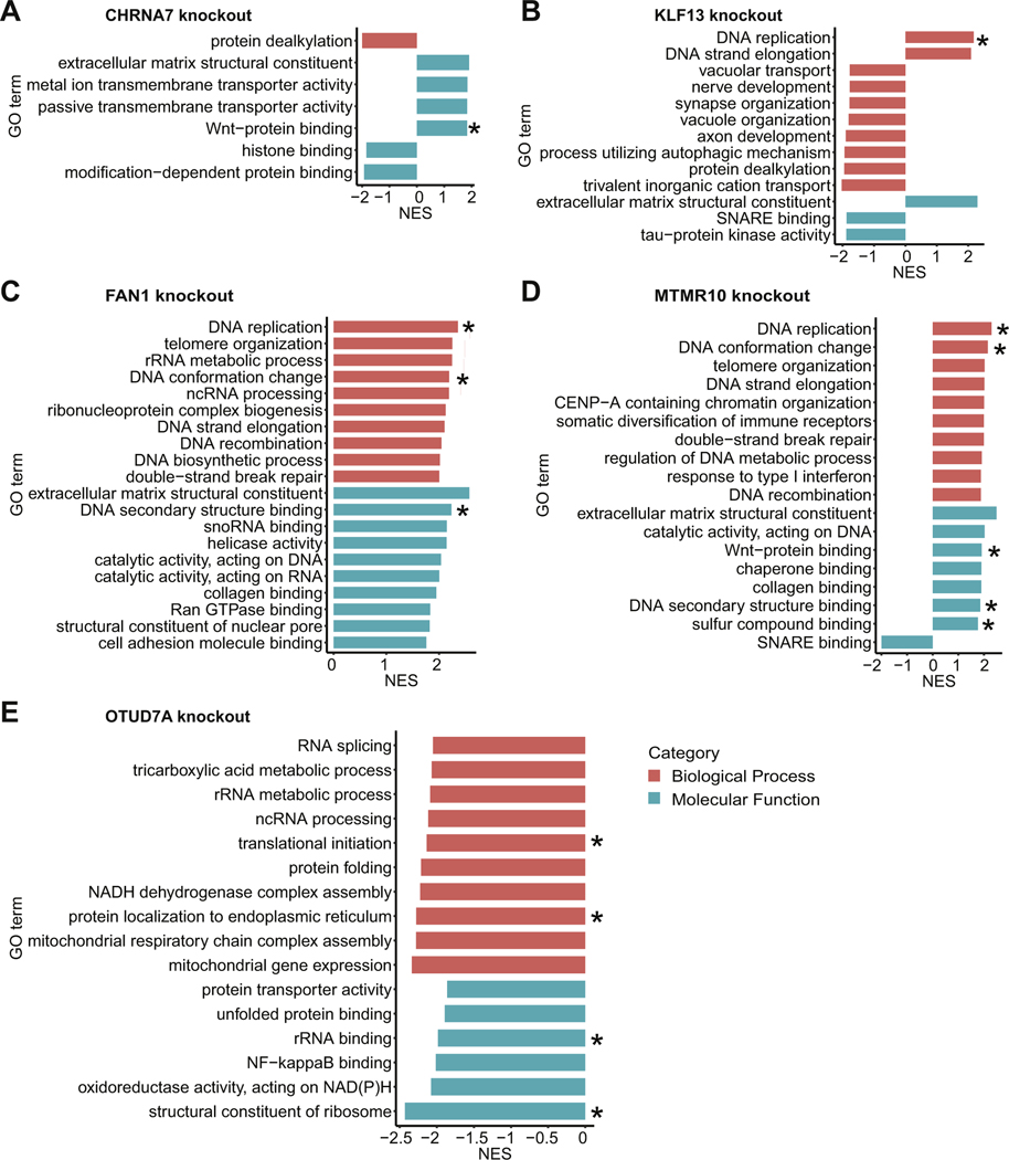
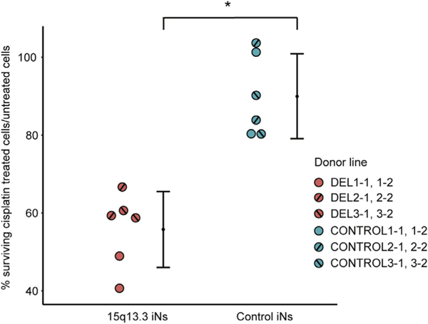
Methods: Induced pluripotent stem cells, from 3 patients with the 15q13.3 microdeletion and 3 control subjects, were generated and converted into induced neurons. We analyzed the effects of the 15q13.3 microdeletion on genome-wide gene expression, DNA methylation, chromatin accessibility, and sensitivity to cisplatin-induced DNA damage. Furthermore, we measured gene expression changes in induced neurons with CRISPR (clustered regularly interspaced short palindromic repeats) knockouts of individual 15q13.3 microdeletion genes.
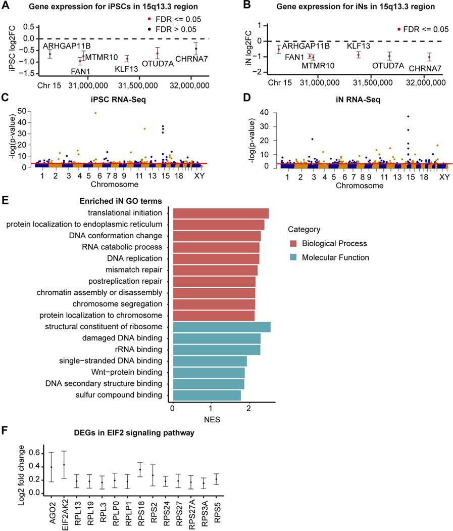
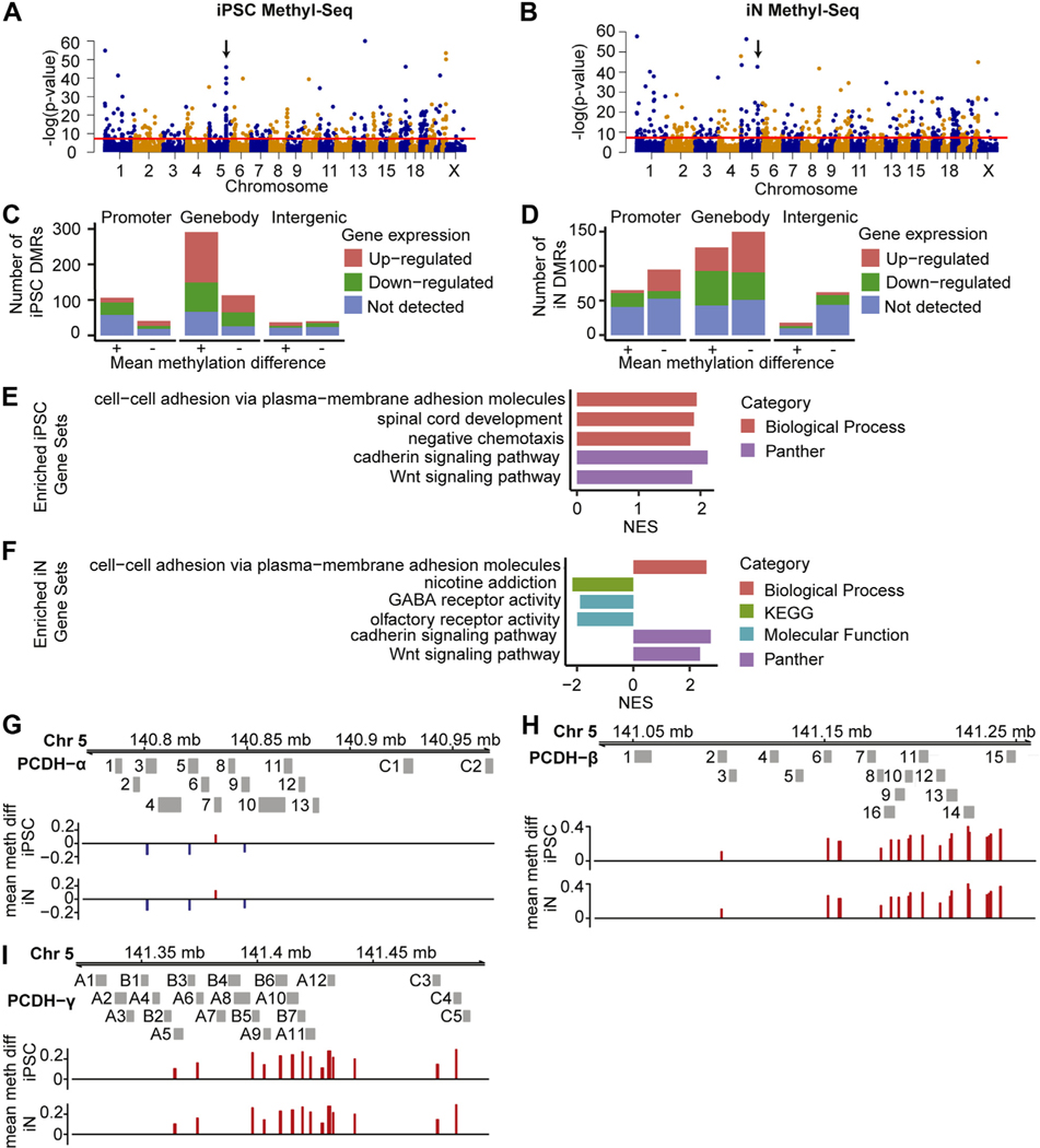
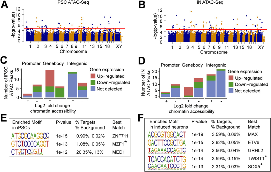
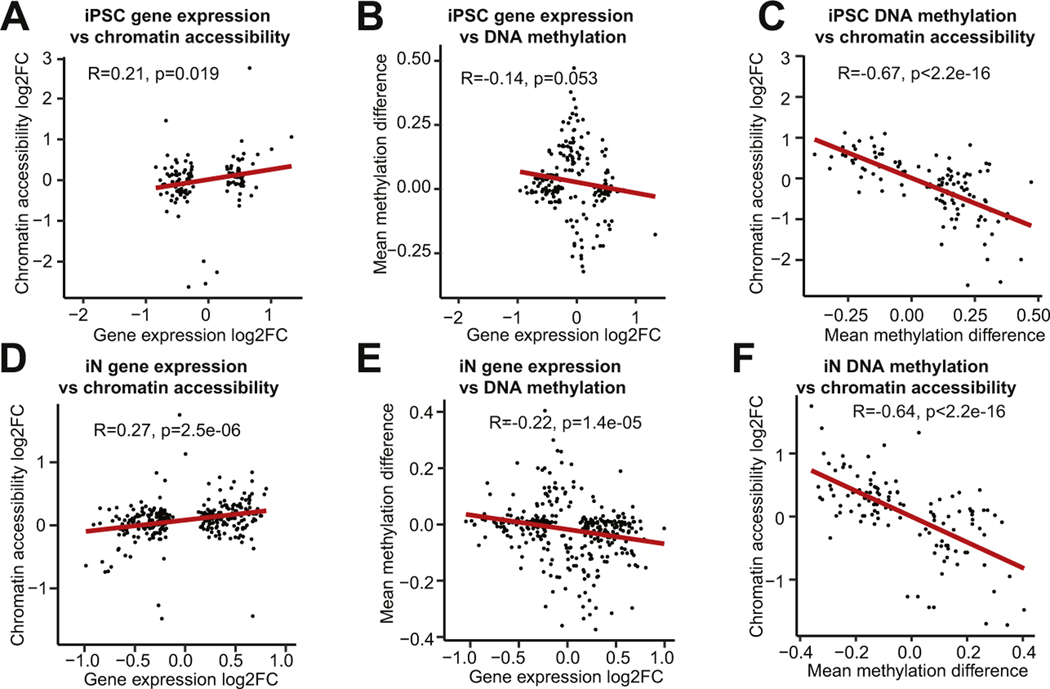
Results: In both induced pluripotent stem cells and induced neurons, gene copy number change within the 15q13.3 microdeletion was accompanied by significantly decreased gene expression and no compensatory changes in DNA methylation or chromatin accessibility, supporting the model that haploinsufficiency of genes within the deleted region drives the disorder. Furthermore, we observed global effects of the microdeletion on the transcriptome and epigenome, with disruptions in several neuropsychiatric disorder-associated pathways and gene families, including Wnt signaling, ribosome function, DNA binding, and clustered protocadherins. Individual gene knockouts mirrored many of the observed changes in an overlapping fashion between knockouts.
Conclusions: Our multiomics analysis of the 15q13.3 microdeletion revealed downstream effects in pathways previously associated with neuropsychiatric disorders and indications of interactions between genes within the deletion. This molecular systems analysis can be applied to other chromosomal aberrations to further our etiological understanding of neuropsychiatric disorders.
Keywords: 15q13.3; CRISPR–Cas9; Copy number variants; Genomics; Induced pluripotent stem cells; Neurons.
Copyright © 2020 Society of Biological Psychiatry. Published by Elsevier Inc. All rights reserved.
Conflict of interest statement
The authors report no biomedical financial interests or potential conflicts of interest.
Figures
Comment in
-
Cross-Platform Validation of 15q13.3 Microdeletion Network Effects in Human Neurons.Biol Psychiatry. 2021 Mar 1;89(5):e25-e27. doi: 10.1016/j.biopsych.2020.12.012. Biol Psychiatry. 2021. PMID: 33541528 No abstract available.
Similar articles
-
Mouse Model of Chromosome 15q13.3 Microdeletion Syndrome Demonstrates Features Related to Autism Spectrum Disorder.J Neurosci. 2015 Dec 9;35(49):16282-94. doi: 10.1523/JNEUROSCI.3967-14.2015. J Neurosci. 2015. PMID: 26658876 Free PMC article.
-
Cross-Platform Validation of 15q13.3 Microdeletion Network Effects in Human Neurons.Biol Psychiatry. 2021 Mar 1;89(5):e25-e27. doi: 10.1016/j.biopsych.2020.12.012. Biol Psychiatry. 2021. PMID: 33541528 No abstract available.
-
OTUD7A Regulates Neurodevelopmental Phenotypes in the 15q13.3 Microdeletion Syndrome.Am J Hum Genet. 2018 Feb 1;102(2):278-295. doi: 10.1016/j.ajhg.2018.01.006. Am J Hum Genet. 2018. PMID: 29395074 Free PMC article.
-
Genetic components of microdeletion syndromes and their role in determining schizophrenia traits.Mol Biol Rep. 2024 Jul 13;51(1):804. doi: 10.1007/s11033-024-09731-y. Mol Biol Rep. 2024. PMID: 39001960 Review.
-
Compound heterozygous microdeletion of chromosome 15q13.3 region in a child with hypotonia, impaired vision, and global developmental delay.Am J Med Genet A. 2014 Jul;164A(7):1815-20. doi: 10.1002/ajmg.a.36535. Epub 2014 Apr 3. Am J Med Genet A. 2014. PMID: 24700535 Review.
Cited by
-
Bioinformatic Evaluation of KLF13 Genetic Variant: Implications for Neurodevelopmental and Psychiatric Symptoms.Genes (Basel). 2024 Aug 11;15(8):1056. doi: 10.3390/genes15081056. Genes (Basel). 2024. PMID: 39202416 Free PMC article.
-
Deepening the understanding of CNVs on chromosome 15q11-13 by using hiPSCs: An overview.Front Cell Dev Biol. 2023 Jan 6;10:1107881. doi: 10.3389/fcell.2022.1107881. eCollection 2022. Front Cell Dev Biol. 2023. PMID: 36684422 Free PMC article. Review.
-
Impaired OTUD7A-dependent Ankyrin regulation mediates neuronal dysfunction in mouse and human models of the 15q13.3 microdeletion syndrome.Mol Psychiatry. 2023 Apr;28(4):1747-1769. doi: 10.1038/s41380-022-01937-5. Epub 2023 Jan 6. Mol Psychiatry. 2023. PMID: 36604605 Free PMC article.
-
Gene Editing in Pluripotent Stem Cells and Their Derived Organoids.Stem Cells Int. 2021 Nov 30;2021:8130828. doi: 10.1155/2021/8130828. eCollection 2021. Stem Cells Int. 2021. PMID: 34887928 Free PMC article. Review.
-
An integrated framework for functional dissection of variable expressivity in genetic disorders.medRxiv [Preprint]. 2025 Jul 24:2025.07.22.25331885. doi: 10.1101/2025.07.22.25331885. medRxiv. 2025. PMID: 40778180 Free PMC article. Preprint.
References
-
- Cubells JF, Deoreo EH, Harvey PD, Garlow SJ, Garber K, Adam MP, et al. (2011): Pharmaco-genetically guided treatment of recurrent rage outbursts in an adult male with 15q13.3 deletion syndrome. Am J Med Genet A 155:805–810. - PubMed
Publication types
MeSH terms
Supplementary concepts
Grants and funding
LinkOut - more resources
Full Text Sources
Other Literature Sources
Molecular Biology Databases
