. 2020 Dec:117:111257.
doi: 10.1016/j.msec.2020.111257. Epub 2020 Jul 6.

Empirical and theoretical insights into the structural effects of selenite doping in hydroxyapatite and the ensuing inhibition of osteoclasts

Affiliations

Empirical and theoretical insights into the structural effects of selenite doping in hydroxyapatite and the ensuing inhibition of osteoclasts

Victoria M Wu et al. Mater Sci Eng C Mater Biol Appl. 2020 Dec.

Abstract

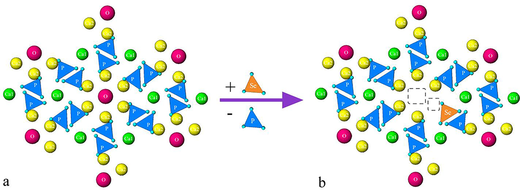
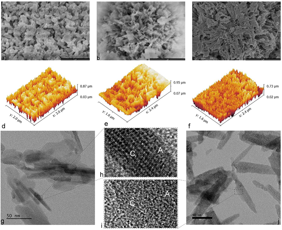
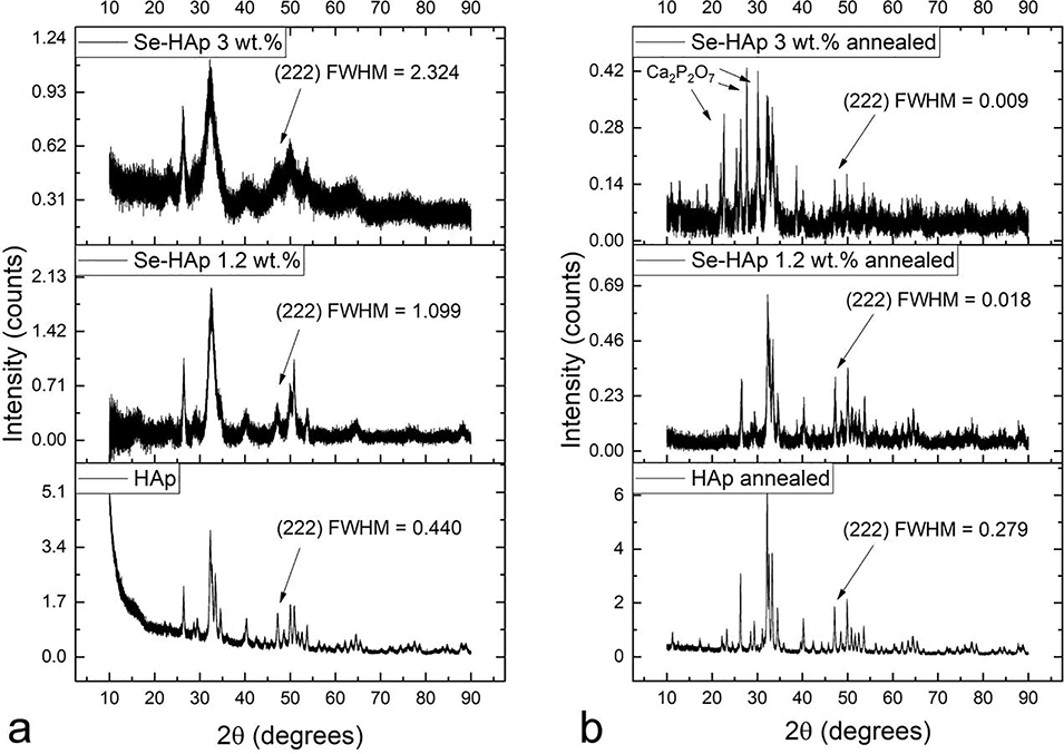
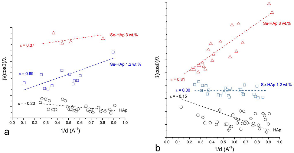
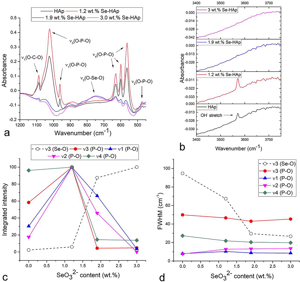
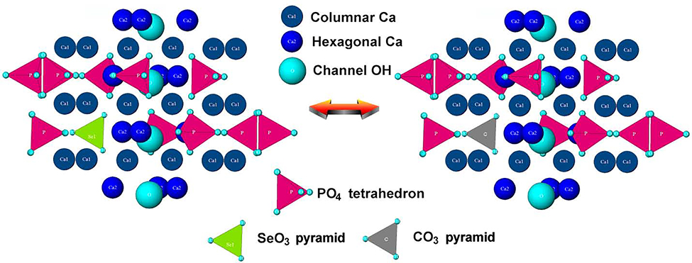
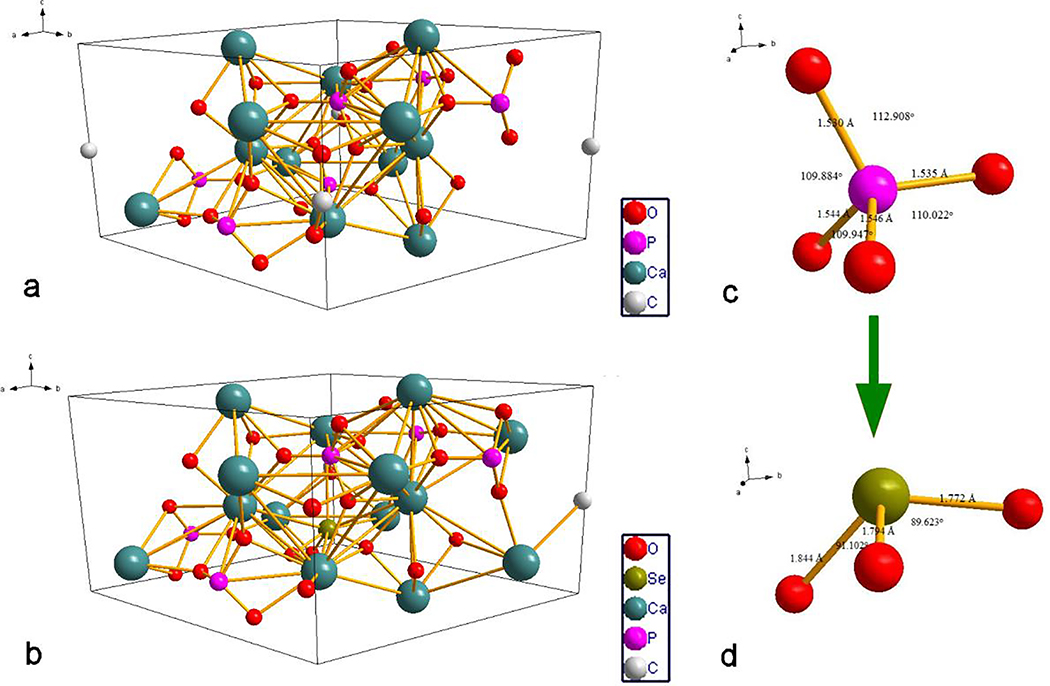
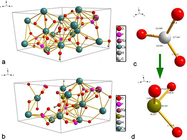
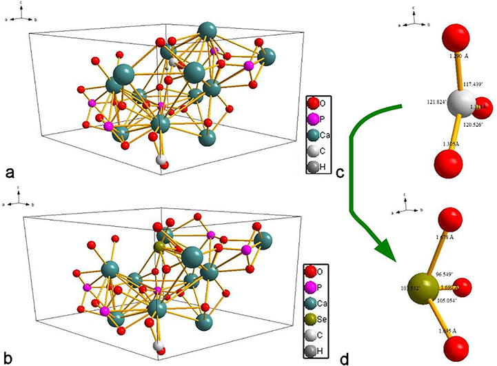
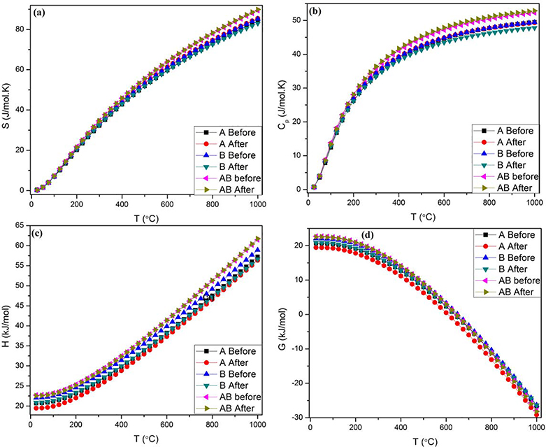
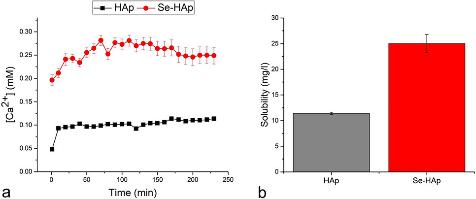
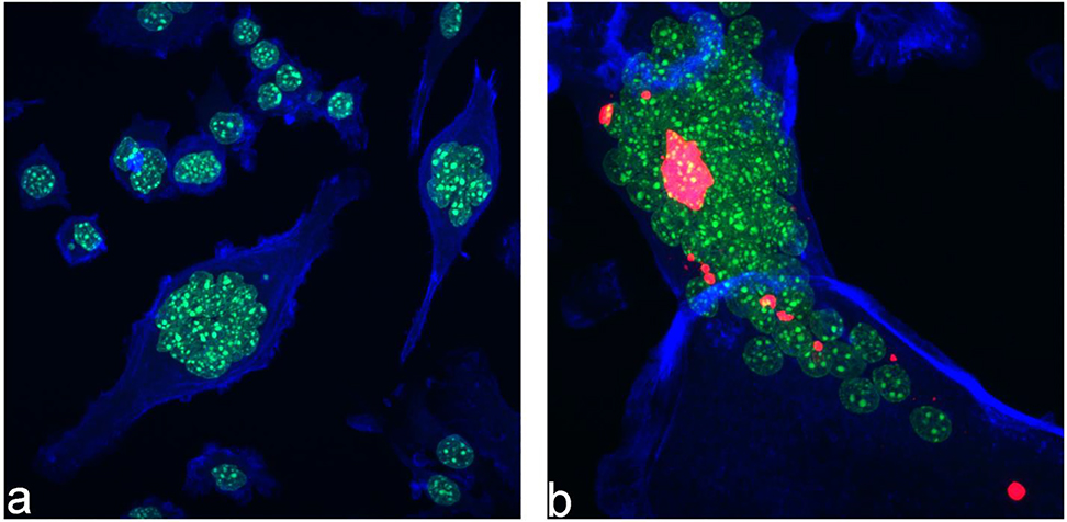
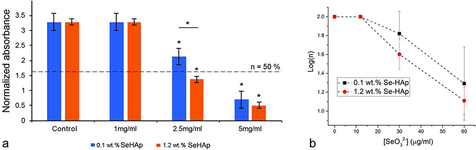
The use of ions as therapeutic agents has the potential to minimize the use of small-molecule drugs and biologics for the same purpose, thus providing a potentially more economic and less adverse means of treating, ameliorating or preventing a number of diseases. Hydroxyapatite (HAp) is a solid compound capable of accommodating foreign ions with a broad range of sizes and charges and its properties can dramatically change with the incorporation of these ionic additives. While most ionic substitutes in HAp have been monatomic cations, their lesser atomic weight, higher diffusivity, chaotropy and a lesser residence time on surfaces theoretically makes them prone to exert a lesser influence on the material/cell interaction than the more kosmotropic oxyanions. Selenite ion as an anionic substitution in HAp was explored in this study for its ability to affect the short-range and the long-range crystalline symmetry and solubility as well as for its ability to affect the osteoclast activity. We combined microstructural, crystallographic and spectroscopic analyses with quantum mechanical calculations to understand the structural effects of doping HAp with selenite. Integration of selenite ions into the crystal structure of HAp elongated the crystals along the c-axis, but isotropically lowered the crystallinity. It also increased the roughness of the material in direct proportion with the content of the selenite dopant, thus having a potentially positive effect on cell adhesion and integration with the host tissue. Selenite in total acted as a crystal structure breaker, but was also able to bring about symmetry at the local and global scales within specific concentration windows, indicating a variety of often mutually antagonistic crystallographic effects that it can induce in a concentration-dependent manner. Experimental determination of the lattice strain coupled with ab initio calculations on three different forms of carbonated HAp (A-type, B-type, AB-type) demonstrated that selenite ions initially substitute carbonates in the crystal structure of carbonated HAp, before substituting phosphates at higher concentrations. The most energetically favored selenite-doped HAp is of AB-type, followed by the B-type and only then by the A-type. This order of stability was entailed by the variation in the geometry and orientation of both the selenite ion and its neighboring phosphates and/or carbonates. The incorporation of selenite in different types of carbonated HAp also caused variations of different thermodynamic parameters, including entropy, enthalpy, heat capacity, and the Gibbs free energy. Solubility of HAp accommodating 1.2 wt% of selenite was 2.5 times higher than that of undoped HAp and the ensuing release of the selenite ion was directly responsible for inhibiting RAW264.7 osteoclasts. Dose-response curves demonstrated that the inhibition of osteoclasts was directly proportional to the concentration of selenite-doped HAp and to the selenite content in it. Meanwhile, selenite-doped HAp had a significantly less adverse effect on osteoblastic K7M2 and MC3T3-E1 cells than on RAW264.7 osteoclasts. The therapeutically promising osteoblast vs. osteoclast selectivity of inhibition was absent when the cells were challenged with undoped HAp, indicating that it is caused by selenite ions in HAp rather than by HAp alone. It is concluded that like three oxygens building the selenite pyramid, the coupling of (1) experimental materials science, (2) quantum mechanical modeling and (3) biological assaying is a triad from which a deeper understanding of ion-doped HAp and other biomaterials can emanate.

Keywords: Ab initio; Calcium phosphate; Hydroxyapatite; Nanoparticles; Osteoblasts; Selenite.


Conflict of interest statement

Declaration of competing interest There are no conflicts of interest to declare.

Figures

Fig.1.
Fig.1.
Crystal structure of pure HAp viewed down the c-axis before (a) and after (b) a single phosphate-to-selenite substitution in the center, showing the two typical vacancies forming to balance the resulting cationic charge excess: Ca2+ and OH. Different ionic elements are represented with different colors: columnar, Ca1 calcium ions with the coordination number of 9 are green; hexagonal, Ca2 calcium ions with the coordination number of 7 are yellow; hydroxyl is magenta; phosphate is blue; selenite is orange.
Fig.2.
Fig.2.
Scanning (a-f) and transmission (g-j) electron micrographs of HAp powders containing 0 (a, d, g, h), 0.1 (b, e) and 1.2 (c, f, i, j) wt.% of selenite ions. Crystalline and amorphous domains in rod-shaped HAp and Se-HAp particles in (e) and (f) are denoted with C and A, respectively.
Fig.3.
Fig.3.
Lattice strain measured as Δc/c and Δa/a in the direction of c- and a- axes, respectively, as a function of the selenite content in co-precipitated and annealed Se-HAp (a) and the average crystallite domain dimensions in the direction of c- and a- axes calculated from the broadening of the halfwidths of (002) (d = 3.440 Å, 2θ = 25.90 °) and (300) (d = 2.720 Å, 2θ = 32.90 °) diffraction peaks, respectively.
Fig.4.
Fig.4.
X-ray diffractograms of HAp and selenite-doped HAp with two different weight contents of selenite, 1.2 and 3.0 wt.%, depending on whether the powders were as-prepared (a) or annealed (b). Full widths at half maxima (FWHM) were measured on the (222) peak at 2θ = 46.69 °, corresponding to the Bragg distance of d = 1.943 Å. Ca2P2O7 denotes calcium pyrophosphate as the secondary phase.
Fig.5.
Fig.5.
Williamson-Hall plots constructed in the 0 – 1 Å−1 range of the reciprocal lattice spacing, d for as-precipitated (a) and annealed (b) HAp powders containing different amounts of selenite: 0, 1.2 or 3.0 wt.%. ε denotes dimensionless lattice strains calculated as halved slopes of the corresponding linear fits.
Fig.6.
Fig.6.
FTIR spectra of HAp powders doped with different concentrations of selenite ion, ranging from 0 to 3 wt.% in the 450 – 1200 cm−1 (a) and 3400 – 3750 cm−1 (b) spectral ranges, along with the integrated intensities (c) and full widths at half maxima (FWHM) (d) of the symmetric, ν1, and asymmetric, ν3, stretching modes and the doubly and triply degenerated, bending modes, ν2 and ν4, respectively, of the phosphate tetrahedron, and the asymmetric, ν3, stretching mode band of the selenite pyramid as a function of the selenite content in HAp.
Fig.7.
Fig.7.
Crystal structure of HAp viewed perpendicularly to the c-axis, showing the competition of selenite and carbonate pyramids for the same phosphate tetrahedron during co-precipitation of Se-HAp under ambient conditions.
Fig.8.
Fig.8.
The preferred geometry for A-type carbonated HAp (a) and the optimum geometry for selenite-doped A-type carbonated HAp (b). Phosphate group orientation with bond lengths and angles in A-type carbonated HAp before doping with selenite (a) and the geometry of the selenite ion in the unit cell of A-type carbonated Se-HAp (d).
Fig.9.
Fig.9.
The preferred geometry for B-type carbonated HAp (a) and the optimum geometry for selenite-doped B-type carbonated HAp (b). Phosphate group orientation with bond lengths and angles in B-type carbonated HAp before doping with selenite (a) and the geometry of the selenite ion in the unit cell of B-type carbonated Se-HAp (d).
Fig.10.
Fig.10.
The preferred geometry for AB-type carbonated HAp (a) and the optimum geometry for selenite-doped AB-type carbonated HAp (b). Phosphate group orientation with bond lengths and angles in AB-type carbonated HAp before doping with selenite (a) and the geometry of the selenite ion in the unit cell of AB-type carbonated Se-HAp (d).
Fig.11.
Fig.11.
Temperature profiles of selected thermodynamic parameters, including entropy (a), heat capacity (b), enthalpy (c) and the Gibbs free energy (d), depending on the type of carbonated HAp before and after the substitution with selenite.
Fig.12.
Fig.12.
Free calcium ion concentration in the aqueous supernatant in a 1 mg/ml suspension (20 mM Bis-Tris, pH 6.8, 25 °C) of as-precipitated, non-annealed HAp, either pure (HAp) or doped with 1.2 wt.% of selenite (Se-HAp) at different time points (a) and the solubility values for the two powders after 24 h of immersion time (b).
Fig.13.
Fig.13.
Fluorescent optical micrographs of control RAW264.7 cells differentiating from mononuclear precursors to polynuclear osteoclastic cells without (a) and with (b) the presence of uptaken Se-HAp particles. Cytoskeletal f-actin microfilaments are stained in blue, cell nuclei in green and Se-HAp particles in red.
Fig.14.
Fig.14.
(a) Absorbance at λ = 540 nm of lysed confluent RAW264.7 osteoclastic cell suspensions normalized to the absorbance of the cell culture medium, indicative of the mitochondrial dehydrogenase activity and, thus, of the cell viability, n, as determined by the MTT assay after 48 h of incubation with different concentrations of Se-HAp particles (1, 2.5 and 5 mg per ml of the cell culture medium) containing different amounts of the selenite ion (0.1 and 1.2 wt.%). Dashed line represents 50 % viability. (b) Logarithmic viability (log(n)) of osteoclastic RAW264.7 cells as a function of the predicted concentration of selenite ions in the medium containing partially dissolved Se-HAp particles with two different selenite contents (0.1 and 1.2 wt.%) and at three different doses (1, 2.5 and 5 mg/ml). Data points are presented as arithmetic means (n = 3) with error bars representing standard deviation. Samples with a significantly lower cell viability with respect to the control group (p < 0.05) are marked with *.
Fig.15.
Fig.15.
Comparative cell viabilities of K7M2 and MC3T3-E1 osteoblastic cells and RAW264.7 osteoclastic cells, as determined by the MTT assay after 48 h of incubation with 5 mg/ml HAp or Se-HAp particles containing different amounts of the selenite ion (0.1 and 1.2 wt.%). Dashed line represents the 100 % viability of the negative control cells not challenged with any particles. Data points are presented as arithmetic means (n = 3) with error bars representing standard deviation. Samples with a significantly lower cell viability with respect to the control group (p < 0.05) are marked with *.

References

    1. Uskoković V, The Role of Hydroxyl Channel in Defining Selected Physicochemical Peculiarities Exhibited by Hydroxyapatite, RSC Adv. 5 (2015) 36614–36633. - PMC - PubMed
    1. Uskoković V, Ion-Doped Hydroxyapatite: An Impasse or the Road to Follow?, Ceram. Int. 46 (2020) 11443–11465.
    1. Mayer I, Berger U, Markitziu A, Gedalia I, The uptake of lithium ions by synthetic carbonated hydroxyapatite, Calcif. Tissue Int. 38 (1986) 293–295. - PubMed
    1. Arslan A, Cakmak S, Gumusderelioglu M, Enhanced osteogenic activity with boron-doped nanohydroxyapatite-loaded poly(butylene adipate-co-terephthalate) fibrous 3D matrix. Artificial Cells, Nanomedicine, and Biotechnology 46 (2018) 790–799. - PubMed
    1. Ciobanu G, Bargan AM, Luca C, New Bismuth-Substituted Hydroxyapatite Nanoparticles for Bone Tissue Engineering. JOM 67 (2015) 2534–42.

LinkOut - more resources