. 2020 Oct:37:101710.
doi: 10.1016/j.redox.2020.101710. Epub 2020 Sep 1.

Vitamin E treatment in NAFLD patients demonstrates that oxidative stress drives steatosis through upregulation of de-novo lipogenesis

Affiliations

Vitamin E treatment in NAFLD patients demonstrates that oxidative stress drives steatosis through upregulation of de-novo lipogenesis

Maren C Podszun et al. Redox Biol. 2020 Oct.

Abstract

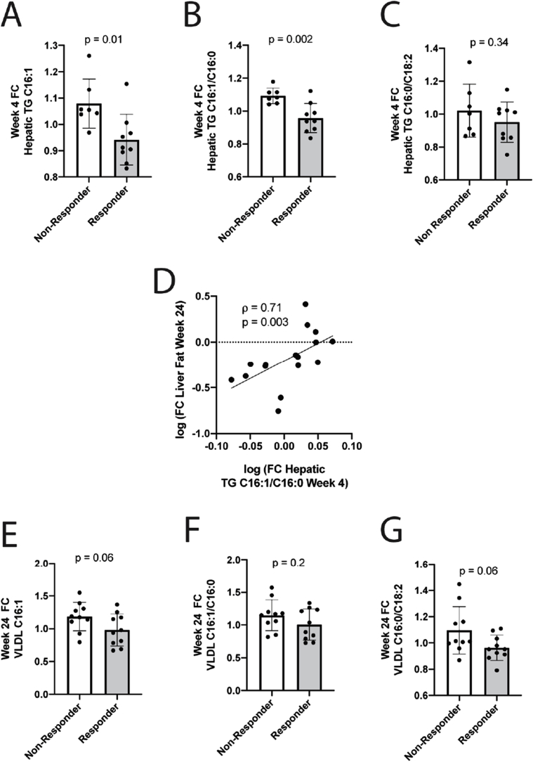
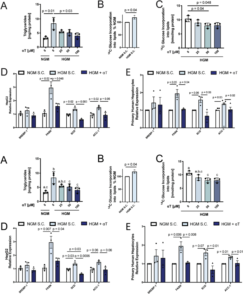
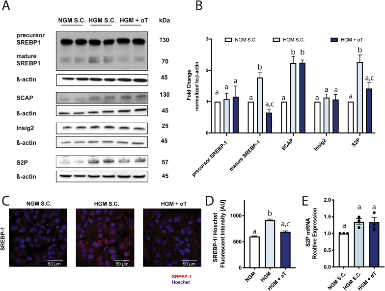
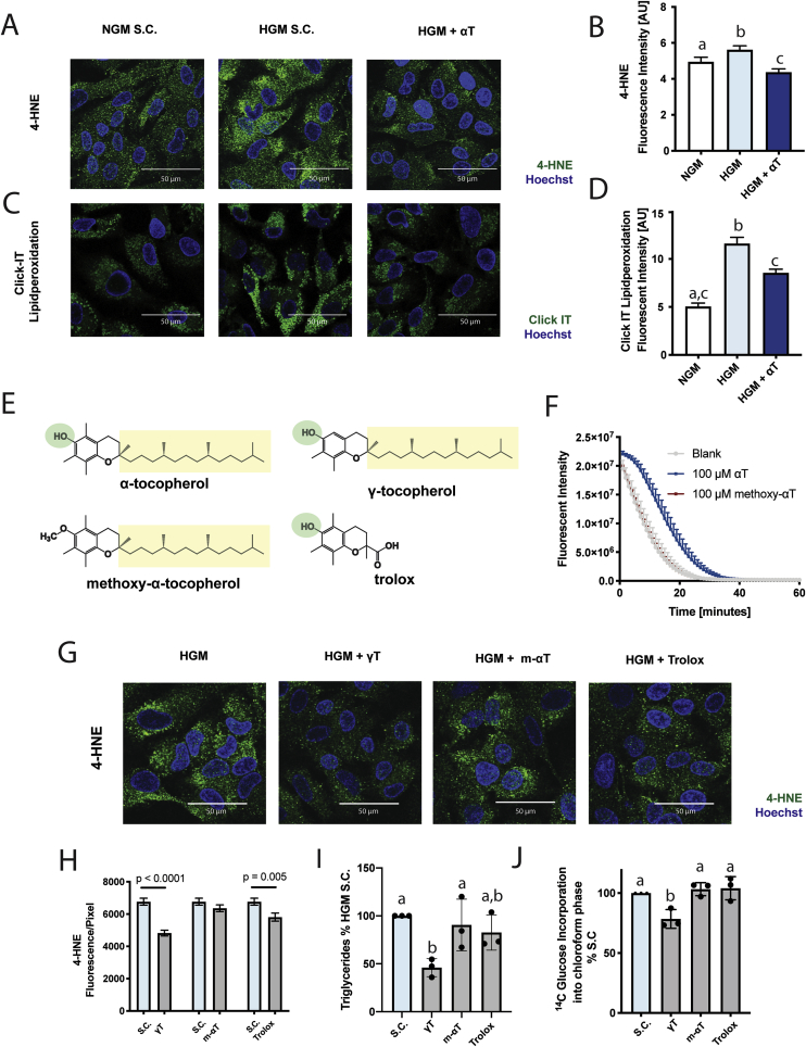
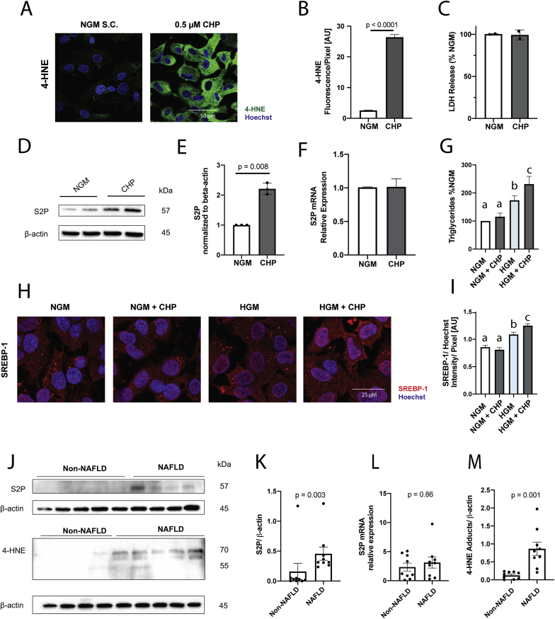
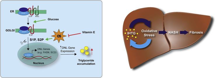
Oxidative stress (OS) in non-alcoholic fatty liver disease (NAFLD) promotes liver injury and inflammation. Treatment with vitamin E (α-tocopherol, αT), a lipid-soluble antioxidant, improves liver injury but also decreases steatosis, thought to be upstream of OS, through an unknown mechanism. To elucidate the mechanism, we combined a mechanistic human trial interrogating pathways of intrahepatic triglyceride (IHTG) accumulation and in vitro experiments. 50% of NAFLD patients (n = 20) treated with αT (200-800 IU/d) for 24 weeks had a ≥ 25% relative decrease in IHTG by magnetic resonance spectroscopy. Paired liver biopsies at baseline and week 4 of treatment revealed a decrease in markers of hepatic de novo lipogenesis (DNL) that strongly predicted week 24 response. In vitro, using HepG2 cells and primary human hepatocytes, αT inhibited glucose-induced DNL by decreasing SREBP-1 processing and lipogenic gene expression. This mechanism is dependent on the antioxidant capacity of αT, as redox-silenced methoxy-αT is unable to inhibit DNL in vitro. OS by itself was sufficient to increase S2P expression in vitro, and S2P is upregulated in NAFLD livers. In summary, we utilized αT to demonstrate a vicious cycle in which NAFLD generates OS, which feeds back to augment DNL and increases steatosis. Clinicaltrials.gov: NCT01792115.

Keywords: NAFLD; Non-alcoholic fatty liver disease; Oxidative stress; S1P; S2P; Vitamin E; de novo lipogenesis.


Conflict of interest statement

The authors have declared no conflict of interest.

Figures

Fig. 1
Fig. 1
αT treatment decreases hepatic DNL in patients with NAFLD. Markers of DNL (C16:1, C16:1/C16:0 and C16:0/C18:2) were quantified in triglycerides from human liver samples before and after 4 weeks of αT treatment (n = 16). Responders (≥25% reduction of IHTG at week 24) had significantly decreased hepatic C16:1 (A) and C16:1/C16:0 ratio (B) and numerically lower C16:0/C18:2 (C). Week 4 changes in hepatic TG C16:1/C16:0 ratio predict change in IHTG content at week 24 (D). DNL markers were measured in VLDL at baseline and week 24 (n = 20). At week 24, responders had a non-significant decrease in VLDL C16:1 (E) and C16:0/C18:2 (G) with little effect on VLDL C16:1/C16:0 (F). A-C, E-G. mean ± SD. Change from baseline to week 4 presented as fold-change (FC). IHTG, intrahepatic triglyceride.
Fig. 2
Fig. 2
αT inhibits de novo lipogenesis (DNL) in vitro. HepG2 cells were incubated with normal growth medium (NGM, white) or high glucose medium (HGM, 25 mmol/L glucose, blue shades) for 48 h. Incorporation of 14C-Glucose into lipids was used as a measure of DNL. Concurrent incubation of cells with 25–100 μmol/L αT decreased cellular triglycerides (A) and DNL (C). αT (100 μmol/L) inhibits HGM-induced upregulation of fatty acid synthase (FASN) and stearoyl-CoA desaturase (SCD) mRNA expression in HepG2 (D) and primary human hepatocytes (PHH, n = 3 donors) (E). PHH experiments were normalized within each donor. All n = 3 experiments; A-C mean ± SD, D-E mean ± SEM. All samples were compared to HGM solvent control (S·C.). Bars not sharing the same letter are significantly different p < 0.05. (For interpretation of the references to colour in this figure legend, the reader is referred to the Web version of this article.)
Fig. 3
Fig. 3
αT decreases SREBP-1 maturation. HepG2 cells incubated with normal growth medium (NGM, white) or high glucose medium (HGM, 25 mmol/L, blue shades) with or without αT (100 μmol/L) for 48 h αT decreased HGM-induced maturation of SREBP-1 without affecting precursor protein levels (A,B) and decreased HGM-induced nuclear translocation (C,D). αT did not affect SCAP or Insig2 (A–B). αT inhibits HGM-driven upregulation S2P at protein (A–B) but not mRNA (E) level. qPCR results were normalized to 18s (E). A - representative Western blot images for respective proteins; B – corresponding quantification. C- representative imaging of nuclear SREBP-1 in cells; D – Corresponding quantification. All data mean ± SEM. B, E n = 3; D n = 2. Solvent Control (S·C.). Bars not sharing the same letter are significantly different p < 0.05. (For interpretation of the references to colour in this figure legend, the reader is referred to the Web version of this article.)
Fig. 4
Fig. 4
αT inhibits de novo lipogenesis through an antioxidant effect. HepG2 cells incubated with normal growth medium (NGM, white) or high glucose medium (HGM, 25 mmol/L, blue shades) with or without αT (100 μmol/L), γ-tocopherol (γT, 75 μmol/L), methoxy-α-tocopherol (m-αT, 100 μmol/L) or trolox (100 μmol/L) for 48 h. HGM significantly induced 4-HNE adduct formation (A, B) and oxidation of ClickIT linoleic acid probe (C, D). Structural formulas of investigated compounds (E). αT, but not m-αT is able to quench oxygen radicals in the ORAC assay (F). γT and trolox inhibit HGM-induced 4-HNE adduct formation, while m-αT does not (G, H). αT and γT are able to decrease glucose-induced triglyceride formation (I) and DNL (J), while m-αT and trolox are ineffective. B, D, H mean ± SEM; I,J mean ± SD. B,D,H n = 2; I-J n = 3. A, C, G show representative images; B, D, I show corresponding quantification. Solvent Control (S·C.). Bars not sharing the same letter are significantly different p < 0.05. (For interpretation of the references to colour in this figure legend, the reader is referred to the Web version of this article.)
Fig. 5
Fig. 5
Oxidative stress increases S2P protein. HepG2 cells were incubated with cumene hydroperoxide (CHP, 0.5 μmol/L, grey) overnight. CHP increased 4-HNE adducts (A,B) without changes in viability (C) and increased S2P protein expression (D,E) without changes in mRNA (F). CHP requires high glucose media (25 mmol/L glucose) to induce triglyceride accumulation (I) and increase SREBP-1 nuclear translocation (G,H). Control (n = 9) and NAFLD (n = 9) liver tissue samples were obtained through the Liver Tissue and Cell Distribution System (LTCDS). NAFLD patients had significantly higher protein levels of S2P (J,K) as well as 4-HNE (M) compared to controls without changes in S2P mRNA (L). qPCR data was normalized to 18s. B,I mean ± SEM, n = 2; C, mean ± SD, n = 2; E,F mean ± SEM, n = 3; G mean ± SD, n = 3; K, L, M mean ± SEM, n = 17. Bars not sharing the same letter are significantly different p < 0.05.
Fig. 6
Fig. 6
Proposed mechanism and NAFLD progression model. (Left) SREBP-1 resides complexed with SCAP and INSIG in the ER. Upon activation, INSIG dissociates and SCAP facilitates transport to Golgi where cleavage by S1P and S2P leads to mature SREBP-1 which translocates to the nucleus and activates lipogenic genes. Vitamin E blocks SREBP-1 translocation through reduced protein expression of S1P and S2P by lowering oxidative stress (OS). (Right) Intra-hepatic triglyceride accumulation (IHTG) induces OS which can lead to progression to non-alcoholic steatohepatitis (NASH) and fibrosis. Our data supports a bi-directional model in which oxidative stress contributes to disease progression and exacerbates IHTG.

References

    1. Younossi Z., Anstee Q.M., Marietti M., Hardy T., Henry L., Eslam M., George J., Bugianesi E. Global burden of NAFLD and NASH: trends, predictions, risk factors and prevention. Nat. Rev. Gastroenterol. Hepatol. 2018;15:11–20. doi: 10.1038/nrgastro.2017.109. - DOI - PubMed
    1. Masarone M., Rosato V., Dallio M., Gravina A.G., Aglitti A., Loguercio C., Federico A., Persico M. Role of oxidative stress in pathophysiology of nonalcoholic fatty liver disease. Oxid. Med. Cell. Longev. 2018:9547613. doi: 10.1155/2018/9547613. (2018) - DOI - PMC - PubMed
    1. Burton G.W., Joyce A., Ingold K.U. First proof that vitamin E is major lipid-soluble, chain-breaking antioxidant in human blood plasma. Lancet (North Am. Ed.) 1982;2:327. - PubMed
    1. Institute of Medicine . Natl. Acad. Press. Washingt. DC; 2000. Dietary Reference Intakes for Vitamin C, Vitamin E, Selenium and Carotenoids; pp. 186–283. - DOI - PubMed
    1. Wang X., Quinn P.J. Vitamin E and its functions in membranes. Prog. Lipid Res. 1999;38:309–336. - PubMed

Publication types

Associated data