

Orbitofrontal sulcogyral morphology in patients with cocaine use disorder
- PMID: 32920245
- PMCID: PMC8126989
- DOI: 10.1016/j.pscychresns.2020.111174
Orbitofrontal sulcogyral morphology in patients with cocaine use disorder
Abstract
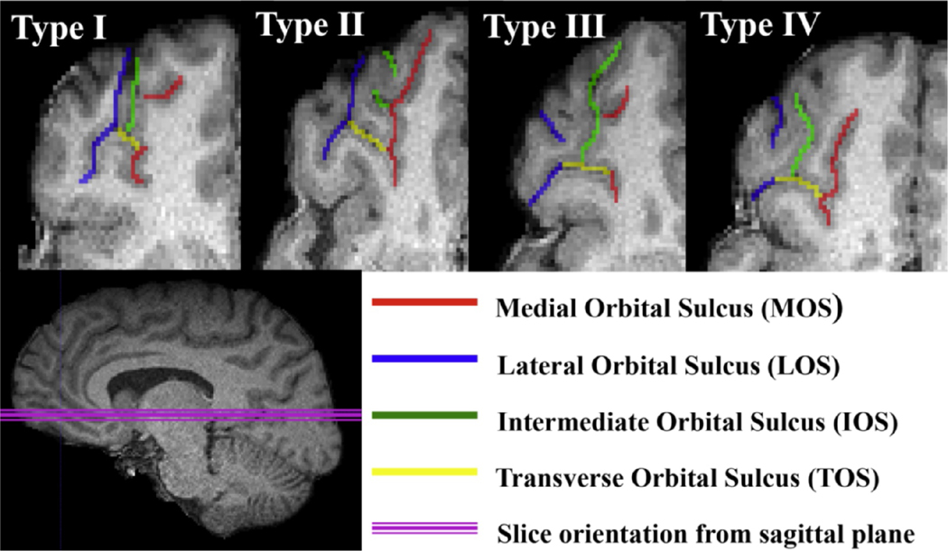
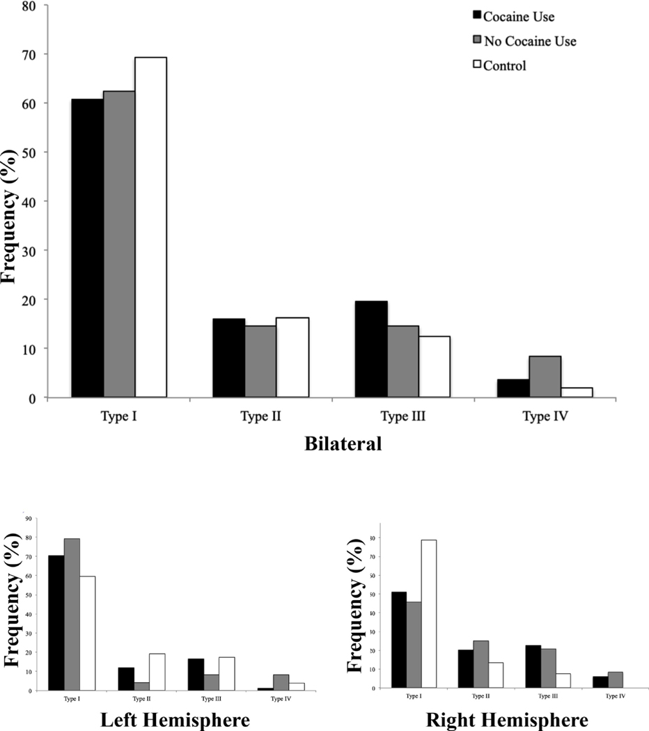
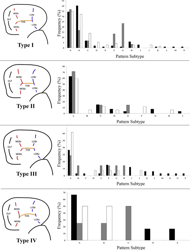
Orbitofrontal cortex (OFC) is thought to be involved in appropriate processing of rewarding stimuli, and abnormal OFC structure and function has been found in patients with substance use disorders. Atypical patterns of the H-sulcus in the OFC have been primarily identified with schizophrenia, but also with bipolar disorder, both of which are associated with comorbid substance use. Given the high rates of substance use within Axis I psychiatric disorders, it is reasonable to consider how frequencies of OFC patterns in populations with only substance use compare to controls. This information is crucial to disentangle whether atypical frequencies of H-sulcus sulcogyral patterns within psychopathology are associated with the psychiatric or substance use phenotype. Here, we present the first analysis of H-sulcus sulcogyral patterns in a population of adult black men with (n = 84) and without (n = 24) cocaine use disorder (CUD). We find that OFC sulcogyral patterns are not significantly different from the control group, indicating that OFC sulcogyral patterns are not disrupted in patients with CUD. As exploratory analyses, we describe OFC sulcogyral pattern subtypes in this cohort as well as an additional control group (n = 52), in order to add to the growing body of literature on OFC sulcogyral pattern characterization.
Keywords: Gyrification; OFC; Sulcus; Ventromedial prefrontal cortex.
Copyright © 2020. Published by Elsevier B.V.
Conflict of interest statement
Declaration of Competing Interest
All authors declare that there are no conflicts of interest.
Figures
Similar articles
-
Orbitofrontal sulcogyral morphology is a transdiagnostic indicator of brain dysfunction.Neuroimage Clin. 2017 Dec 18;17:910-917. doi: 10.1016/j.nicl.2017.12.021. eCollection 2018. Neuroimage Clin. 2017. PMID: 29527495 Free PMC article.
-
Orbitofrontal sulcogyral pattern and olfactory sulcus depth in the schizophrenia spectrum.Eur Arch Psychiatry Clin Neurosci. 2016 Feb;266(1):15-23. doi: 10.1007/s00406-015-0587-z. Epub 2015 Mar 11. Eur Arch Psychiatry Clin Neurosci. 2016. PMID: 25757375
-
An evaluation of automated tracing for orbitofrontal cortex sulcogyral pattern typing.J Neurosci Methods. 2019 Oct 1;326:108386. doi: 10.1016/j.jneumeth.2019.108386. Epub 2019 Aug 1. J Neurosci Methods. 2019. PMID: 31377175 Free PMC article.
-
Effects of NRG1 genotypes on orbitofrontal sulcogyral patterns in Japanese patients diagnosed with schizophrenia.Psychiatry Clin Neurosci. 2016 Jul;70(7):261-8. doi: 10.1111/pcn.12384. Epub 2016 Apr 8. Psychiatry Clin Neurosci. 2016. PMID: 26909665
-
Orbitofrontal Sulcogyral Pattern as a Transdiagnostic Trait Marker of Early Neurodevelopment in the Social Brain.Clin EEG Neurosci. 2020 Jul;51(4):275-284. doi: 10.1177/1550059420904180. Epub 2020 Feb 6. Clin EEG Neurosci. 2020. PMID: 32028799 Free PMC article. Review.
Cited by
-
Genetics and prescription opioid use (GaPO): study design for consenting a cohort from an existing biobank to identify clinical and genetic factors influencing prescription opioid use and abuse.BMC Med Genomics. 2021 Oct 26;14(1):253. doi: 10.1186/s12920-021-01100-z. BMC Med Genomics. 2021. PMID: 34702274 Free PMC article.
-
Patterns and predictors of orbitofrontal sulcogyral morphology in a nonclinical population.Imaging Neurosci (Camb). 2024 Dec 19;2:imag-2-00389. doi: 10.1162/imag_a_00389. eCollection 2024. Imaging Neurosci (Camb). 2024. PMID: 40800483 Free PMC article.
-
Towards Deciphering the Fetal Foundation of Normal Cognition and Cognitive Symptoms From Sulcation of the Cortex.Front Neuroanat. 2021 Sep 28;15:712862. doi: 10.3389/fnana.2021.712862. eCollection 2021. Front Neuroanat. 2021. PMID: 34650408 Free PMC article.
References
-
- Center for Behavrioral Health Statistics and Quality (CBHSQ), 2015. National survey on drug use and health: detailed tables. Substance Abuse and Mental Health Services Administration. Rockville, MD: 2016.
-
- Centers for Disease Control and Prevention (CDC), Center for Behavioral Health Statistics and Quality, 2015. Behavioral Health Trends in the United States: Results from the 2014. National Survey on Drug Use and Health.
Publication types
MeSH terms
Substances
Grants and funding
LinkOut - more resources
Full Text Sources
Medical
Research Materials
