. 2020 Oct:60:102989.
doi: 10.1016/j.ebiom.2020.102989. Epub 2020 Sep 10.

Cell motility and migration as determinants of stem cell efficacy

Affiliations

Cell motility and migration as determinants of stem cell efficacy

Lusine Danielyan et al. EBioMedicine. 2020 Oct.

Abstract

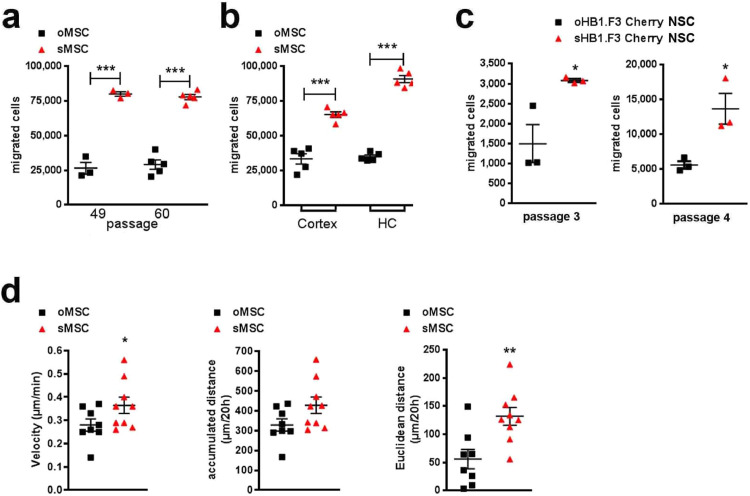
Background: Stem cells` (SC) functional heterogeneity and its poorly understood aetiology impedes clinical development of cell-based therapies in regenerative medicine and oncology. Recent studies suggest a strong correlation between the SC migration potential and their therapeutic efficacy in humans. Designating SC migration as a denominator of functional SC heterogeneity, we sought to identify highly migrating subpopulations within different SC classes and evaluate their therapeutic properties in comparison to the parental non-selected cells.

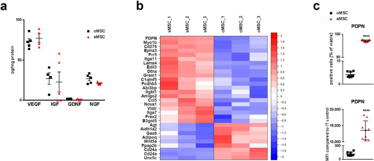
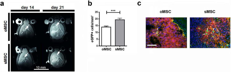
Methods: We selected highly migrating subpopulations from mesenchymal and neural SC (sMSC and sNSC), characterized their features including but not limited to migratory potential, trophic factor release and transcriptomic signature. To assess lesion-targeted migration and therapeutic properties of isolated subpopulations in vivo, surgical transplantation and intranasal administration of MSCs in mouse models of glioblastoma and Alzheimer's disease respectively were performed.

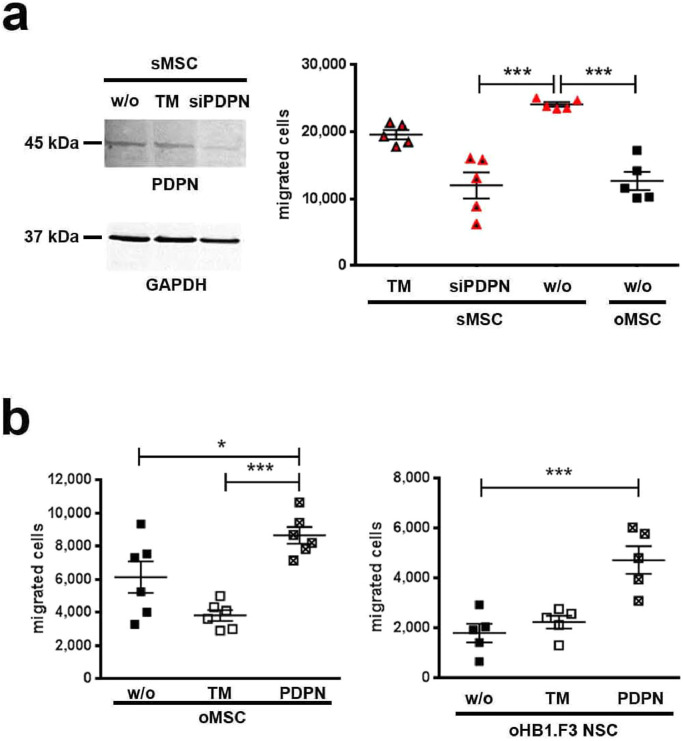
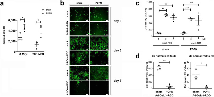
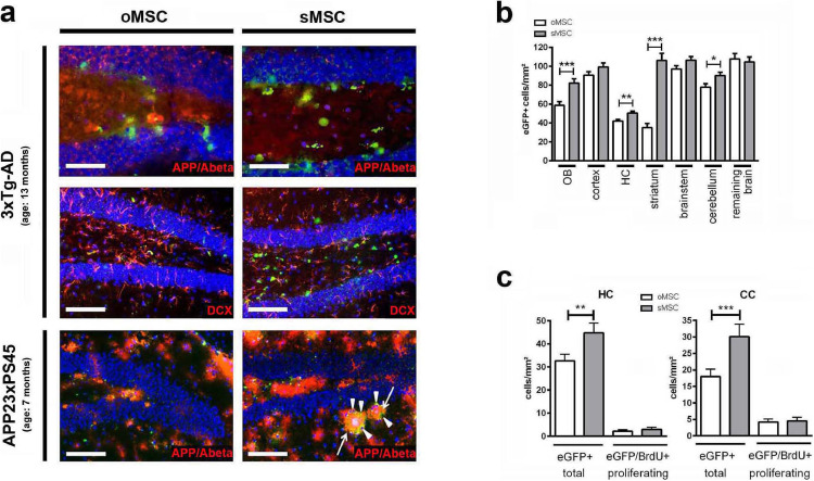
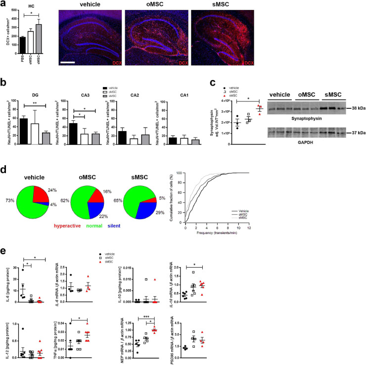
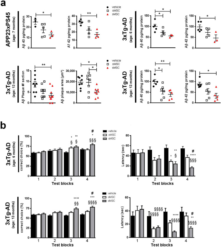
Findings: Comparison of parental non-selected cells with isolated subpopulations revealed superior motility and migratory potential of sMSC and sNSC in vitro. We identified podoplanin as a major regulator of migratory features of sMSC/sNSC. Podoplanin engineering improved oncovirolytic activity of virus-loaded NSC on distantly located glioblastoma cells. Finally, sMSC displayed more targeted migration to the tumour site in a mouse glioblastoma model and remarkably higher potency to reduce pathological hallmarks and memory deficits in transgenic Alzheimer's disease mice.

Interpretation: Functional heterogeneity of SC is associated with their motility and migration potential which can serve as predictors of SC therapeutic efficacy.

Funding: This work was supported in part by the Robert Bosch Stiftung (Stuttgart, Germany) and by the IZEPHA grant.

Keywords: Alzheimer´s disease; Glioblastoma; Intranasal; Mesenchymal stem cells; Neural stem cells; Oncovirolysis.


Conflict of interest statement

Declaration of Competing Interest Dr. Danielyan reports grants from IZEPHA, during the conduct of the study. Prof.. Schwab, Dr. Schaeffeler and Dr. Winter report grants from Robert Bosch Stiftung (Stuttgart, Germany), during the conduct of the study. Dr. Danielyan, Dr. Schäfer, Prof. Gleiter and Prof. Schwab have a patent US14/634,501, US14/634,484, PCT/EP2016/054055, DE102012107879, EP2888348 pending. All other authors have no competing interests.

Figures

Fig 1
Fig. 1
In vitro migration potential, characterization and function of murine MSC and human NSC subpopulations (a) Migration capacity of oMSC and sMSC over 3 h at two passages (P49 and P60), n=3-6, each data point represents one biological replicate per group; exemplary experiment of 8 independent experiments; cell numbers of migrated oMSC vs. sMSC in two different passages were compared with ANOVA and Bonferroni's multiple comparisons test (***p <0.001). Error bars: SEM. (b) Migration of oMSC and sMSC over 3 h towards murine cortical (cortex) and hippocampal (HC) primary cultures seeded on the bottom of transwell chambers, n=5, each data point represents one biological replicate per group; cell numbers of oMSC vs. sMSC migrated towards cortical and hippocampal cultures were compared with ANOVA and Bonferroni's multiple comparisons test (***p <0.001). Error bars: SEM. (c) Migration over 4.5 h of human o/sHB1.F3 Cherry NSC in passage 3 and 4. The number of floating cells in the lower compartment of the migration chamber was counted after crystal violet staining. Cumulative cell number calculated from 79 optical fields within a 24-well plate, n=3 per passage, each data point represents one biological replicate per group out of a single experiment. Cell numbers of migrated oNSC vs. sNSC were compared with two-tailed t-test (*p<0.05); Error bars: SEM. (d) Two hours after seeding (100,000 cells/19.6 cm2), oMSC and sMSC were tracked every 30 min during 20 h. Velocity, accumulated and Euclidian distance for representative n=9 cells were determined by manual tracking. Each data point represents a single cell analysed per group. Two-tailed t-test was used to compare the velocity, accumulated and Euclidian distance of oMSC vs. sMSC (*p<0.05; **p<0.01); Error bars: SEM.
Fig 2
Fig. 2
In vitro paracrine properties and transcriptomic signature of oMSC and sMSC (a) Secreted trophic factors (mRNA clustered in Supplementary Fig.2a) in cell culture supernatants of oMSC and sMSC 48 h after plating of cells analysed by ELISA, n=5, each data point represents one biological replicate per group; replicates analysed collectively by specific ELISAs for GDNF, IGF, NGF and VEGF; oMSC were compared with sMSC by ANOVA with Bonferroni's multiple comparisons test. Error bars: SEM; GDNF, Glial cell-derived neurotrophic factor; IGF, Insulin-like growth factor; NGF, Nerve growth factor; VEGF, vascular endothelial growth factor. (b) Heatmap of microarray data displaying genes related to cell migration and/or adhesion. Only genes differing significantly and relevantly between murine oMSC and sMSC are displayed. Row-wise z-scores of normalized log2 signal intensities in murine oMSC and sMSC, n=3 per group (3 different passages). Z-scores color-coded as indicated by colour bar on the right. (c) Quantification of PDPN protein by flow cytometry analysis of o/sMSC to validate PDPN mRNA expression (microarray), n=9, each data point represents one biological replicate per group; two-tailed t-test was used for comparison of percentage and MFI counts of oMSC vs. sMSC (***p<0.001); Error bars: SD. IT, isotype control; MFI, mean fluorescence intensity.
Fig 3
Fig. 3
In vitro loss and gain of PDPN function in MSC and NSC. (a) PDPN expression in sMSC was suppressed by siRNA (siPDPN). Shown by Western Blot. Migration was analysed for untransfected (w/o) sMSC, sMSC incubated with transfection medium (TM) only, siPDPN transfected sMSC, and non-transfected (w/o) oMSC (migration time: 3 h), n=5, each data point represents one biological replicate per group out of a single experiment; ANOVA and Bonferroni's multiple comparisons test was used to compare non-transfected (w/o) oMSC with siPDPN transfected or untransfected sMSC, (***p <0.001). Error bars: SEM. (b) Migration over 4 h of untransfected (w/o) oMSC/oNSC, oMSC/oNSC incubated with TM only, and PDPN overexpressing oMSC/oNSC was analysed after 120 h (oMSC) or 72 h (oNSC) after PDPN transfection, n=6 (oMSC), n=5 (oNSC), each data point represents one biological replicate per group; ANOVA and Bonferroni's multiple comparisons test was used to compare PDPN transfected cells with TM-treated or untransfected (w/o) oMSC/oNSC (*p<0.05; ***p<0.001). Error bars: SEM.
Fig 4
Fig. 4
In vitro glioma tropism and oncovirolytic properties of selected and non-selected human NSC (a) oHB1.F3 NSC transfected with PDPN or treated with transfection reagent (sham) were subsequently infected with 200 MOI of Ad-Delo3-RGD to induce oncovirolysis of glioma cells or left untreated (0 MOI; mock) for 2 h, followed by a 4 h migration period through 8 µm transwell membranes towards the conditioned medium from LNT-229 glioma cells. Migrated cells were harvested and counted. Number of migrated mock- or Ad-Delo3-RGD infected F3 NSC after 4h of migration was compared between PDPN-transfected vs. untransfected cells by two-tailed t-test (*p<0.05), n=3, each data point represents one biological replicate per group; MOI, multiplicity of infection; Error bars: SEM. (b) Migrated NSC with and without PDPN overexpression were co-cultured with eGFP-labelled LNT-229 glioma cells. Representative microphotographs from co-cultures taken at the start (day 0), at day 5 and at day 7 of co-culture. Scale bars: 60 µm. (c) Growth of glioma cells in presence of migrated mock-NSC was quantified as described in methods. Cell density of glioma cells (normalized to the cell density at day 0) in the presence of mock-NSC with and without PDPN transfection was analysed by ANOVA and Bonferroni's multiple comparisons test (*p<0.05), n=3, each data point represents one biological replicate per group out of a single experiment; Error bars: SEM. (d) Growth of glioma cells in the presence of migrated Ad-Delo3-RGD infected F3 NSC. Cell density of glioma cells at day 5 and day 7 is normalized to that of day 0. For comparison of sham- vs. PDPN-transfected NSC two-tailed t-test (*p<0.05) was used, n=3, each data point represents one biological replicate per group; MOI, multiplicity of infection; Error bars: SEM.
Fig 5
Fig. 5
Localisation and quantification of o/sMSC in the brain of AD mouse models (a) Appearance of eGFP+ oMSC and sMSC (green) in the dentate gyri of 13-month-old 3xTg-AD mice (two upper panels) and 7-month-old APP23xPS45 mice (lower panel). Both animal models show strong expression of APP/Amyloid beta (red). eGFP+ oMSC and sMSC detected in vicinity to DCX positive neurons (red) and circumvent (arrowheads) Aβ plaques (arrows). Scale bars: 100 µm, except for the two microphotographs on the top (50 µm). INA, intranasal application; DCX, doublecortin. (b) Quantification of eGFP+ oMSC and sMSC in different brain regions of 7-month-old 3xTg-AD mice (n=6 per group) after intranasal application (INA) of eGFP+ o/sMSC (2 x application of 1 × 106 cells). 20 sections from each mouse brain were analysed. oMSC and sMSC cell counts normalized to mm² were compared in each brain region by two-tailed t-test (*p <0.05; **p <0.01; ***p<0.001). Each column represents ≥60 data points obtained from 20 sections (3-10 micrographs per section); Error bars: SEM. HC, hippocampus; OB, olfactory bulb. (c) Quantification of total eGFP+ o/sMSC and proliferating eGFP/BrdU+ o/sMSC in the HC and CC of 13-month-old 3xTg-AD mice (n=4 per group) after INA. Total o/sMSC were quantified (total oMSC vs. total sMSC) and also compared for their positivity for BrdU (eGFP/BrdU+ oMSC vs. sMSC) from 25 brain sections from each animal by ANOVA and Bonferroni's multiple comparisons test (**p <0.01; ***p <0.001). Each column represents ≥25 data points obtained from 25 sections (1-2 micrographs per section). Error bars: SEM. CC, corpus callosum; HC, hippocampus. (For interpretation of the references to color in this figure legend, the reader is referred to the web version of this article.)
Fig 6
Fig. 6
Neuroprotective and neurogenesis inducing features of oMSC and sMSC in transgenic AD mice (a) Photomicrographs of DCX expression and quantification of DCX+ neurons in the hippocampi of 13-month-old 3xTg-AD mice after INA of o/sMSC (2 x application of 1 × 106 cells, n=3 per group). The number of DCX+ cells was quantified from 10 brain sections of each animal and normalized to mm². Each column represents 9-10 data points obtained from 10 sections. ANOVA with Bonferroni's multiple comparisons test was used to compare vehicle-, oMSC and sMSC-treated groups (*p <0.05). Error bars: SEM. Scale bar: 500 µm. DCX, doublecortin. (b) Quantification of NeuN/TUNEL+ neurons in the DG and CA1-3 in 13-month-old 3xTg-AD mice (n=4 per group) after INA of oMSC and sMSC. Cells were quantified in 10 sections from each brain and normalized to mm². Each column represents 6-11 data points obtained from 10 sections. ANOVA with Bonferroni's multiple comparisons test was used to compare vehicle-, oMSC and sMSC-treated groups (*p <0.05; **p <0.01). Error bars: SEM. CA, cornu ammonis; DG, dentate gyrus. (c) Western blot with densitometric analysis of synaptophysin in brain homogenates of 13-month-old 3xTg-AD mice (n=3 per group) after INA of oMSC and sMSC. ANOVA with Bonferroni's multiple comparisons test was used to compare vehicle-, oMSC and sMSC-treated groups (*p <0.05). Error bars: SEM. (d) In vivo activity of layer 2/3 neurons located in the vicinity of amyloid plaques in the cortex of 7-8-month-old APP23xPS45 mice after INA of vehicle or o/sMSC (2x application of 1 × 106 cells, n=5 mice per group). Left panel: Neurons classified based on the frequency of their spontaneous Ca2+ transients as silent (0-0.25 transients/min), normal (0.26-4 transients/min), and hyperactive (>4 transients/min) . Pie charts show fractions of hyperactive (red), normal (green) and silent (blue) cells. The relative fractions of different cell types are significantly different (Chi-square test. Vehicle vs. oMSC: p=0.0006, ChiSquare=14.95, df=2; vehicle vs. sMSC: p<0.0001, ChiSquare=31.83, df=2; oMSC vs. sMSC: p=0.034, ChiSquare=6.79, df=2). Right panel: Cumulative histogram showing frequency distribution of Ca2+ transients recorded from neurons close to amyloid plaques after INA of vehicle (225 cells), oMSC (285 cells), and sMSC (310 cells). oMSC and sMSC treatment shifted the frequency distribution significantly towards lower values, and therefore reduced pathological hyperactivity in cortical neurons (Kolmogorov-Smirnov-test. Vehicle vs. oMSC: p<0.0001, D=0.204; Vehicle vs. sMSC: p<0.0001, D=0.336; oMSC vs. sMSC: p=0.0002, D=0.173). (e) Analysis of indicated protein levels by V-plex® analysis (n=5-8 per group; individual samples were collectively analysed by V-plex®) and mRNA levels by qPCR (n=4-5 per group; individual samples collectively analysed for the respective targets) after INA of oMSC and sMSC. IL-12 and IFNγ were barely detectable (data not shown). ANOVA with Bonferroni's multiple comparisons test was used to compare vehicle-, oMSC and sMSC-treated groups (*p <0.05; ***p <0.001). Error bars: SEM. IL, interleukin; NEP, neprilysin. (For interpretation of the references to color in this figure legend, the reader is referred to the web version of this article.)
Fig 7
Fig. 7
Aβ pathology and memory deficits after intranasal oMSC or sMSC treatment in transgenic AD mice (a) Human Aβ 40 and 42 quantified by ELISA in 7-month-old APP23/PS45 mice (n=4 per group), 6-month-old 3xTg-AD mice (n=5 per group) and 13-months-old 3xTg-AD mice (n=5 per group) after INA of o/sMSC (2x application of 1 × 106 cells). Aβ plaque number and cumulative area in the hippocampi of 13-month-old 3xTg-AD mice (n=3 per group) after INA of o/sMSC (2x application of 1 × 106 cells). Number of plaques and cumulative area per brain section quantified from 9-10 brain sections per animal using the APP/Aβ antibody 6E10, each data point represents plaque number or plaque area calculated per section. Each column represents 8-10 data points obtained from 9-10 sections. To compare vehicle-, oMSC and sMSC-treated groups ANOVA with Bonferroni's multiple comparisons test was used (*p <0.05; **p <0.01). Error bars: SEM. (b) Performance in T-maze of 6- and 13-month-old 3xTg-AD mice (n=8 per group) prior (test block 1) and after (test blocks 2-4) INA of o/s MSC (2x application of 1 × 106 cells) shown in test blocks with 4 consecutive test days in each block. Experiments performed once with 6-month-old mice, and twice in 13-month-old mice. The test blocks 2-4 were performed from day 13 to day 28 after INA. In the “correct choice” assessment, each bar represents 32 values of correct choice percentages out of 6 runs on 4 consecutive days of one test block assessed from 8 animals per group. In the latency assessment each bar represents 24-42 data points of latency to reach the correct arm on the last day of each test block from 6-8 mice out of 8 mice analysed. #-comparison between oMSC and sMSC, *-comparison between o/sMSC and PBS (vehicle) group, §-comparison of the respective treatment group in test blocks after INA vs. test block 1 (for instance oMSC of test block 1 vs. oMSC in test blocks 2-4). ANOVA with Bonferroni's multiple comparisons test was used (*p <0.05; **p <0.01; ***p<0.001). Error bars: SEM.
Fig 8
Fig. 8
In vivo intracerebral administration of oMSC and sMSC in an orthotopic model of glioblastoma (a) GL-261-Luc (1 × 105 cells) were injected into the brain of C57BL/6 mice and a T2-weighted MR-anatomy was acquired 14 and 21 days post injection. eGFP+ oMSC or sMSC (1 × 105 cells) were injected into the contralateral side on day 15 after intracerebral injection of GL-261-Luc cells. Tumours are indicated with arrows. (b) Quantification of eGFP+ oMSC and sMSC in the tumour area 7 days after intracerebral injection of eGFP-MSC (n=7 per group). The columns represent 150-180 data points obtained from 20-25 sections per mouse brain. The numbers of eGFP+ oMSC and sMSC were compared by two-tailed t-test (***p <0.001); Error bars: SEM. (c) eGFP+ oMSC and sMSC (green) highlighted with arrows in the GL-261-Luc (red) tumour area 7 days after intracerebral injection of 1 × 105 eGFP+ o/sMSC into the contralateral hemisphere. Scale bar: 100 µm. (For interpretation of the references to color in this figure legend, the reader is referred to the web version of this article.)

Similar articles

Cited by

References

    1. Fu Liu, Halim Ju, Luo Song. Mesenchymal stem cell migration and tissue repair. Cells. 2019;8:784. - PMC - PubMed
    1. Mendonça LS, Onofre I, Miranda CO, Perfeito R, Nóbrega C, de Almeida LP. Stem cell-based therapies for polyglutamine diseases. Adv. Exp. Med. Biol. 2018;1049:439–466. - PubMed
    1. Terunuma A, Ashiba K, Takane T, Sakaguchi Y, Terunuma H. Comparative transcriptomic analysis of human mesenchymal stem cells derived from dental pulp and adipose tissues. J Stem Cells Regen Med. 2019;15:8–11. - PMC - PubMed
    1. Severe N, Karabacak NM, Gustafsson K, Baryawno N, Courties G, Kfoury Y. Stress-induced changes in bone marrow stromal cell populations revealed through single-cell protein expression mapping. Cell Stem Cell. 2019;25:570–583.e7. - PMC - PubMed
    1. Majore I, Moretti P, Hass R, Kasper C. Identification of subpopulations in mesenchymal stem cell-like cultures from human umbilical cord. Cell Commun Signal. 2009;7 - PMC - PubMed

MeSH terms