. 2020 Sep 12;12(17):17738-17753.
doi: 10.18632/aging.103955. Epub 2020 Sep 12.

Anti-aging effects of Ribes meyeri anthocyanins on neural stem cells and aging mice

Affiliations

Anti-aging effects of Ribes meyeri anthocyanins on neural stem cells and aging mice

Jiaming Gao et al. Aging (Albany NY). .

Abstract

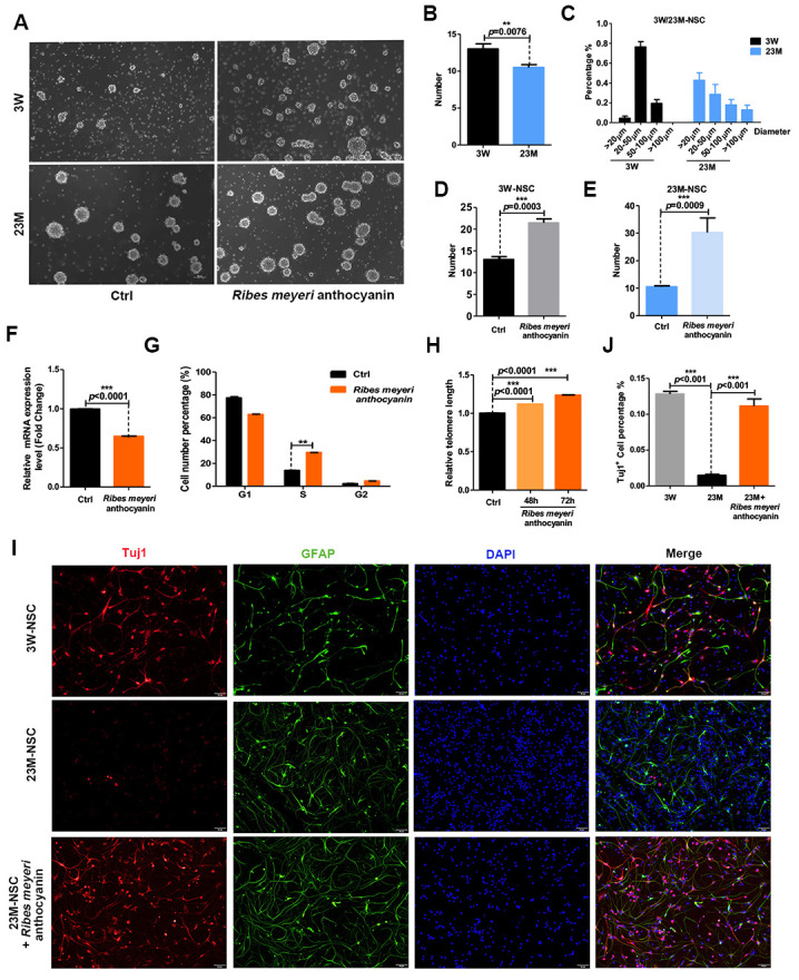
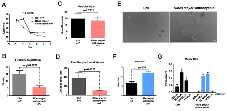
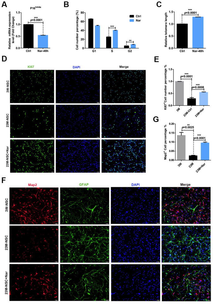
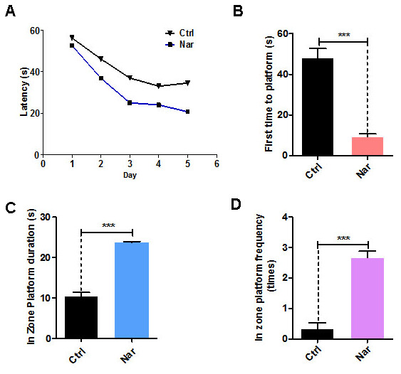
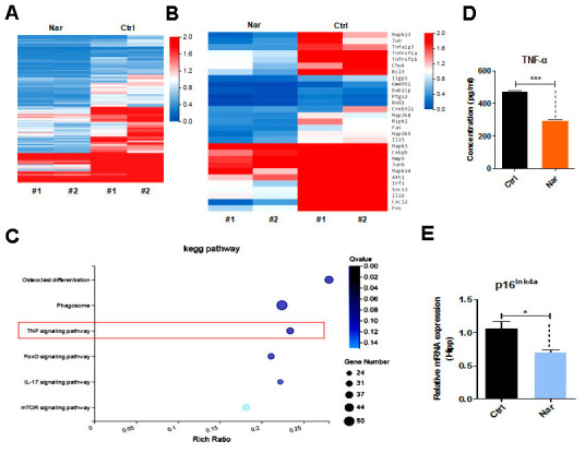
Aging is associated with neurological impairment and cognitive decline. Flavonoids are very promising in anti-aging research in mouse models. Ribes meyeri anthocyanins are rich in abundant flavonoids, but their anti-aging biological activities remain unknown. In this study, we prepared an R. meyeri anthocyanin extract and analyzed its effects on neural stem cell (NSC) senescence in vivo and in vitro. We isolated mouse NSCs and used cell counting kit-8 (CCK-8), cell cycle, reactive oxygen species (ROS), and immunofluorescence methods to analyze the anti-aging effects of R. meyeri anthocyanins as well as naringenin (Nar), which metabolic analysis revealed as an important flavonoid in R. meyeri anthocyanins. RNA-sequencing (RNA-seq) and enzyme-linked immuno sorbent assay (ELISA) methods were also used to investigate Nar-specific mechanisms of anti-aging. After R. meyeri anthocyanin treatment, NSC proliferation accelerated, and NSCs had decreased senescence markers, and reduced P16ink4a expression. R. meyeri anthocyanin treatment also reversed age-dependent neuronal loss in vivo and in vitro. Nar blocked mNSC aging in vitro and improved spatial memory and cognitive abilities in aging mice through downregulation of plasma TNF-α protein. These findings suggest that R. meyeri anthocyanins increase NSC proliferation and improve neurogenesis with aging via Nar-induced reductions in TNF-α protein levels in vivo.

Keywords: Ribes meyeri anthocyanin; aging; cognition; naringenin; senescence.


Conflict of interest statement

CONFLICTS OF INTEREST: All authors declare that no conflicts of interest exists.

Figures

Figure 1
Figure 1
Ribes meyeri anthocyanins rescue the senescence phenotype of mouse neural stem cells (mNSCs). (A) Neurospheres clonally derived from 23-month-old (23M-NSC) and 3-week-old (3W-NSC) mouse brains and treated with 100 pg/mL R. meyeri anthocyanins. (B) Differences in neurosphere numbers between 23M-NSC and 3W-NSC. (C) The shape of 3W-NSC neurospheres was more uniform, with an increased number and size (20–50 μm) compared with 23M-NSC neurospheres. (D, E) Treatment with R. meyeri anthocyanins increased the numbers of both 3W-NSC- and 23M-NSC-derived neurospheres compared with controls. (FH) In 23M-NSCs treated with 100 pg/mL R. meyeri anthocyanins for 48 h, p16ink4a mRNA expression, cell cycle, and relative telomere lengths were measured. (F) Cell senescence marker p16ink4a mRNA expression detected by qRT-PCR. (G) Cell cycles were determined using flow cytometry. (H) qRT-PCR detection of relative telomere length. (I) Immunofluorescence staining of TuJ1 and GFAP in 23M-NSCs treated with R. meyeri anthocyanins and control. (J) Quantification of (I). Data are presented as the mean ± SD of three independent experiments. *P < 0.05, **P < 0.01, and ***P < 0.0001 compared with untreated cells.
Figure 2
Figure 2
Ribes meyeri anthocyanins enhance mouse memory and learning abilities by increasing neural stem cells (NSCs). (A) Time taken to find the platform. (B) Time taken to first reach the platform. (C) Mean velocity to find the platform. (D) Distance swum before finding the platform. (E, F) Treatment with R. meyeri anthocyanins increased neurosphere number and size compared with controls. (G) Neurospheres of 50–100 μm in size were increased in number by R. meyeri anthocyanin treatment in mice. Data are presented as the mean ± SD of three independent experiments. *P < 0.05, **P < 0.01, and ***P < 0.0001 compared with untreated cells. “n” indicates the number of animals in each experimental group.
Figure 3
Figure 3
Effect of naringenin (Nar) on senescence of mouse neural stem cells (mNSCs). (A) p16ink4a mRNA expression was measured by qRT-PCR in 23M-NSCs with and without treatment of 6.8 μg/mL Nar for 48 h. (B) Cell cycle phase distributions of 23M-NSCs treated with Nar for 48 h and control cells. (C) The relative telomere length of 23M-NSCs increased significantly with Nar treatment. (D) Immunofluorescence Ki67 staining of mNSCs treated with Nar for 48 h, with DAPI nuclear labeling. (E) Quantification of (D). (F) Representative fields of MAP2 and GFAP immunofluorescence staining of cultured mNSCs after control and Nar treatment. (G) Quantification of (F). Data are presented as the mean ± SD of three independent experiments. *P < 0.05, **P < 0.01, and ***P < 0.0001 compared with untreated cells.
Figure 4
Figure 4
Naringenin (Nar) enhances memory and learning abilities in aging mice. (A) Escape latency of Nar-treated aging mice was shorter compared with the control group. (B) Time taken to first reach the platform was shorter in Nar-treated aging mice than in the control group. (C) Duration of time spent in the platform zone. (D) Frequency of being observed in the platform zone for Nar-treated aging mice compared with the control group. Data are presented as the mean ± SD of three independent experiments. *P < 0.05, **P < 0.01, and, ***P < 0.0001 compared with the control group. “n” indicates the number of animals used for each experimental group.
Figure 5
Figure 5
Tumor necrosis factor (TNF)-α plays an essential role in naringenin (Nar)-mediated anti-aging effects. (A) Summary of gene expressions in mouse blood, as determined by RNA-seq. (B) Heatmap showing differentially expressed genes (DEGs) in the blood between control and Nar-treated mice. DEGs were identified by a fold change of > 2.0 or < 0.5. (C) Bar graph showing the major correlated signaling pathways. (D) Heatmap showing DEGs in the TNF signaling pathway (E) Plasma TNF-α levels determined by enzyme-linked immunosorbent assay. (E) p16ink4a gene expression levels, as determined by qRT-PCR. Data are presented as the mean ± SD of three independent experiments. *P < 0.05, **P < 0.01, and ***P < 0.0001 compared with the control group.

References

    1. Lu L. A study on the genus Ribes L. in China. Acta Phytotaxonomica Sinica. 1995; 33:58–75.
    1. Xiu JC, Hui Z, Zhao WG, Zhang XY, Sheng C. Studies on Development and Utilization of Libes Resources in Area of Changbai Mountains. 2002; 24:75–74. 10.1088/1009-1963/11/5/313 - DOI
    1. Jing L, Liang A, Yuhong L, Junzhen C. Study on the chemical constituents and the anti-inflammation effects of the leaves of Ribes meyeri. West China Journal of Pharmaceutical Sciences. 2018.
    1. Zhang D, Yuan W, Ma M, Liu M, University Q. Genetic Differentiation Study of Chloroplast DNA trnL-F in Ribes meyeri Maxim. from Qinghai. Genomics & Applied Biology; 2014.
    1. Zhang GL. Assay Study on Total Flavanoids in Chabiaozi(Ribes mandshuricum Komarov). Food Science. 2007.