

lncRNA PART1 and MIR17HG as ΔNp63α direct targets regulate tumor progression of cervical squamous cell carcinoma
- PMID: 32920922
- PMCID: PMC7648017
- DOI: 10.1111/cas.14649
lncRNA PART1 and MIR17HG as ΔNp63α direct targets regulate tumor progression of cervical squamous cell carcinoma
Abstract
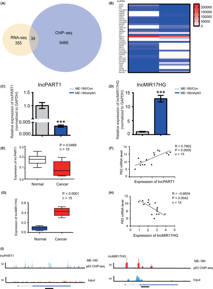
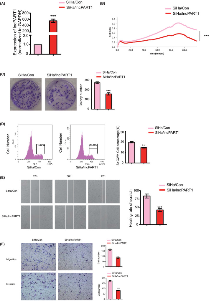
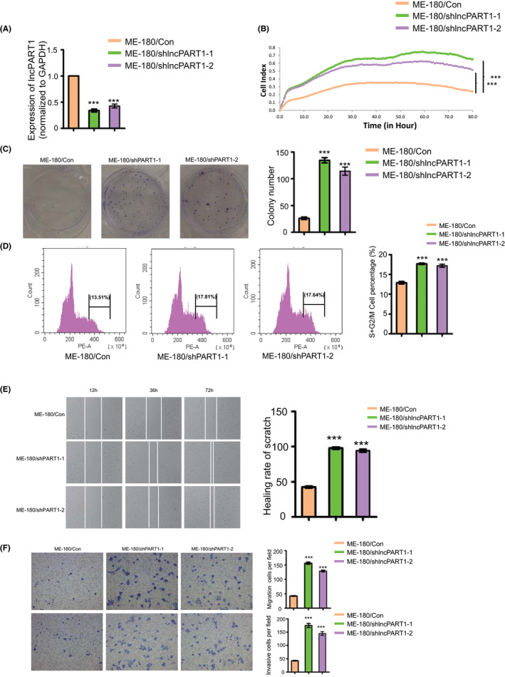
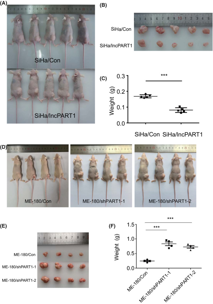
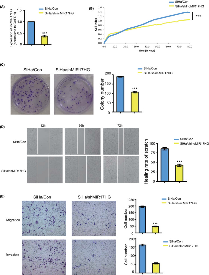
Cervical cancer (CC) remains one of the leading causes of mortality of female cancers worldwide, with more than 90% being cervical squamous cell carcinoma (CSCC). ΔNp63α is the predominant isoform expressed in cervical epithelial tissues and exerts its antitumor function in CSCC. In this study, we have identified 39 long noncoding RNAs as ΔNp63α targets in CSCC through RNA sequencing and chromatin immunoprecipitation sequencing, in which we further confirmed and focused on the two tumor-related long noncoding RNAs, PART1 (lncPART1) and MIR17HG (lncMIR17HG). Experiments from stable overexpression/knockdown cell lines revealed that lncPART1 and lncMIR17HG regulated cell proliferation, migration, and invasion. In vivo experiments further showed that lncPART1 suppresses tumor growth in CSCC-derived tumors. Examinations of clinical tissues indicated that the expression of lncPART1 was positively correlated with ΔNp63α expression, while lncMIR17HG was negatively correlated with ΔNp63α expression, suggesting that ΔNp63α plays a central role via regulating its direct targets in the progression of CSCC. These findings provide novel insights in targeted therapy of cervical cancers.
Keywords: CSCC; cervical cancer; lncMIR17HG; lncPART1; ΔNp63α.
© 2020 The Authors. Cancer Science published by John Wiley & Sons Australia, Ltd on behalf of Japanese Cancer Association.
Conflict of interest statement
The authors have no conflict of interest to declare. All authors have read the journal's authorship agreement. The manuscript has been reviewed by and approved by all named authors.
Figures
Similar articles
-
LncRNA NEF inhibits migration and invasion of HPV-negative cervical squamous cell carcinoma by inhibiting TGF-β pathway.Biosci Rep. 2019 Apr 26;39(4):BSR20180878. doi: 10.1042/BSR20180878. Print 2019 Apr 30. Biosci Rep. 2019. PMID: 30910843 Free PMC article.
-
Down-regulation of lncRNA snaR is correlated with postoperative distant recurrence of HPV-negative cervical squamous cell carcinoma.Biosci Rep. 2018 Nov 13;38(6):BSR20181213. doi: 10.1042/BSR20181213. Print 2018 Dec 21. Biosci Rep. 2018. PMID: 30249756 Free PMC article.
-
ΔNp63α exerts antitumor functions in cervical squamous cell carcinoma.Oncogene. 2020 Jan;39(4):905-921. doi: 10.1038/s41388-019-1033-x. Epub 2019 Oct 1. Oncogene. 2020. PMID: 31576015
-
LncRNA ANCR downregulates hypoxia‑inducible factor 1α and inhibits the growth of HPV‑negative cervical squamous cell carcinoma under hypoxic conditions.Mol Med Rep. 2020 Jan;21(1):413-419. doi: 10.3892/mmr.2019.10792. Epub 2019 Nov 5. Mol Med Rep. 2020. PMID: 31746351
-
Cervical carcinoma high expressed 1 (CCHE1): An oncogenic lncRNA in diverse neoplasms.Biomed Pharmacother. 2021 Oct;142:112003. doi: 10.1016/j.biopha.2021.112003. Epub 2021 Aug 9. Biomed Pharmacother. 2021. PMID: 34385101 Review.
Cited by
-
LncRNA miR-17-92a-1 cluster host gene (MIR17HG) promotes neuronal damage and microglial activation by targeting the microRNA-153-3p/alpha-synuclein axis in Parkinson's disease.Bioengineered. 2022 Feb;13(2):4493-4516. doi: 10.1080/21655979.2022.2033409. Bioengineered. 2022. PMID: 35137671 Free PMC article.
-
Long non-coding RNA MIR17HG sponges microRNA-21 to upregulate PTEN and regulate homoharringtonine-based chemoresistance of acute myeloid leukemia cells.Oncol Lett. 2022 Jan;23(1):24. doi: 10.3892/ol.2021.13142. Epub 2021 Nov 18. Oncol Lett. 2022. PMID: 34868361 Free PMC article.
-
Identification and verification of the role of crucial genes through which methionine restriction inhibits the progression of colon cancer cells.Oncol Lett. 2022 Jun 22;24(2):274. doi: 10.3892/ol.2022.13394. eCollection 2022 Aug. Oncol Lett. 2022. PMID: 35782898 Free PMC article.
-
Long non‑coding RNA PART1: dual role in cancer.Hum Cell. 2022 Sep;35(5):1364-1374. doi: 10.1007/s13577-022-00752-y. Epub 2022 Jul 21. Hum Cell. 2022. PMID: 35864416 Review.
-
Circular RNA ARHGAP5 inhibits cisplatin resistance in cervical squamous cell carcinoma by interacting with AUF1.Cancer Sci. 2023 Apr;114(4):1582-1595. doi: 10.1111/cas.15723. Epub 2023 Feb 2. Cancer Sci. 2023. PMID: 36632741 Free PMC article.
References
-
- Song W, Wang J, Liu H, et al. Effects of LncRNA Lnc‐LIF‐AS on cell proliferation, migration and invasion in a human cervical cancer cell line. Cytokine. 2019;120:165‐175. - PubMed
-
- Aalijahan H, Ghorbian S. Long non‐coding RNAs and cervical cancer. Exp Mol Pathol. 2019;106:7‐16. - PubMed
-
- Siegel RL, Miller KD, Jemal A. Cancer statistics, 2018. CA Cancer J Clin. 2018;68:7‐30. - PubMed
-
- Rodriguez‐Carunchio L, Soveral I, Steenbergen RD, et al. HPV‐negative carcinoma of the uterine cervix: a distinct type of cervical cancer with poor prognosis. BJOG. 2015;122:119‐127. - PubMed
