Review
. 2021 Jan;19(1):7-19.
doi: 10.1111/jth.15097. Epub 2020 Oct 25.

Novel blood coagulation molecules: Skeletal muscle myosin and cardiac myosin

Affiliations
Review

Novel blood coagulation molecules: Skeletal muscle myosin and cardiac myosin

Hiroshi Deguchi et al. J Thromb Haemost. 2021 Jan.

Abstract

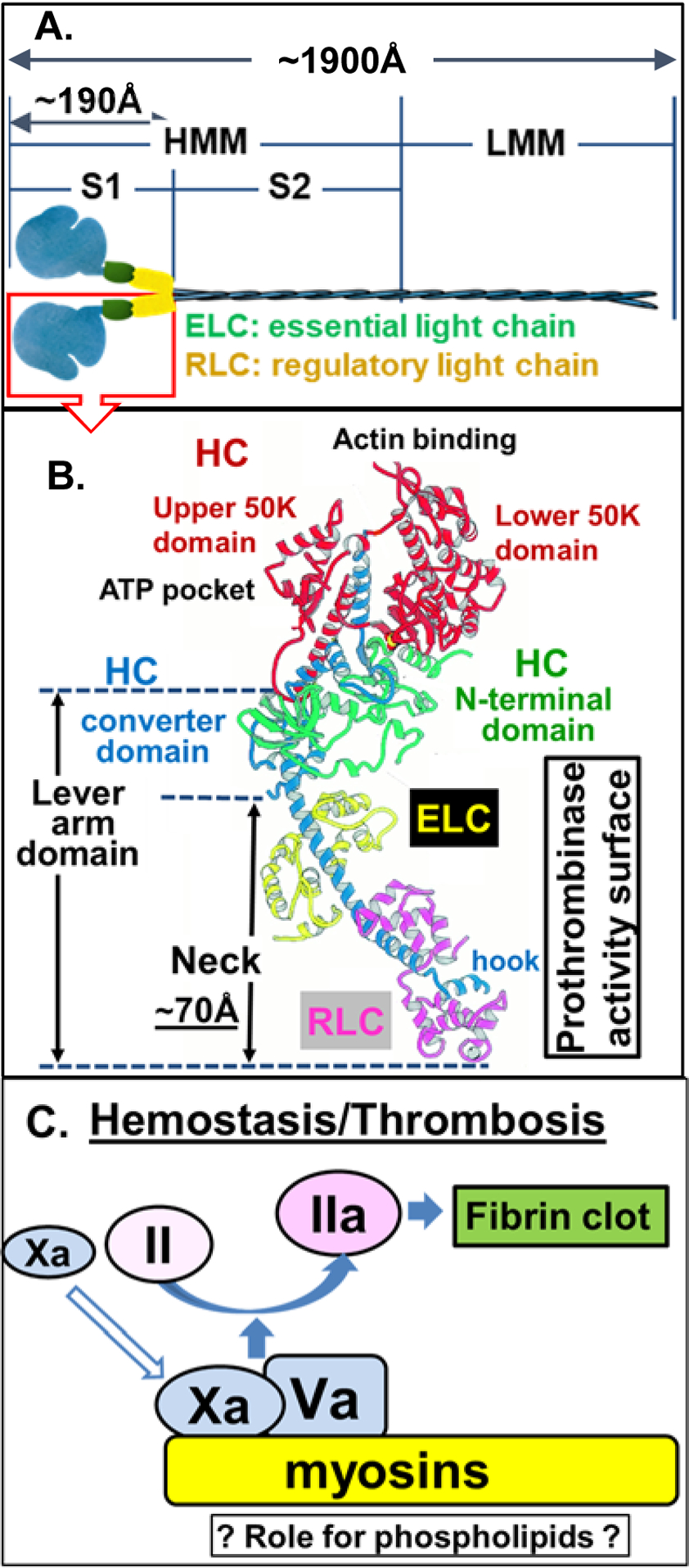
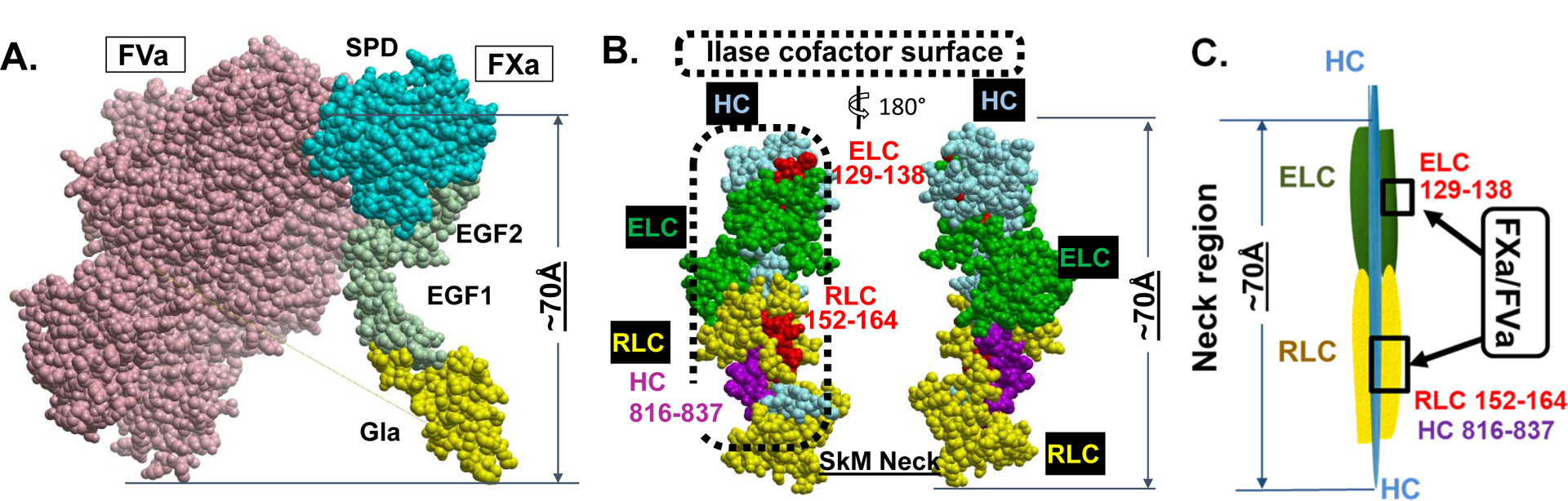
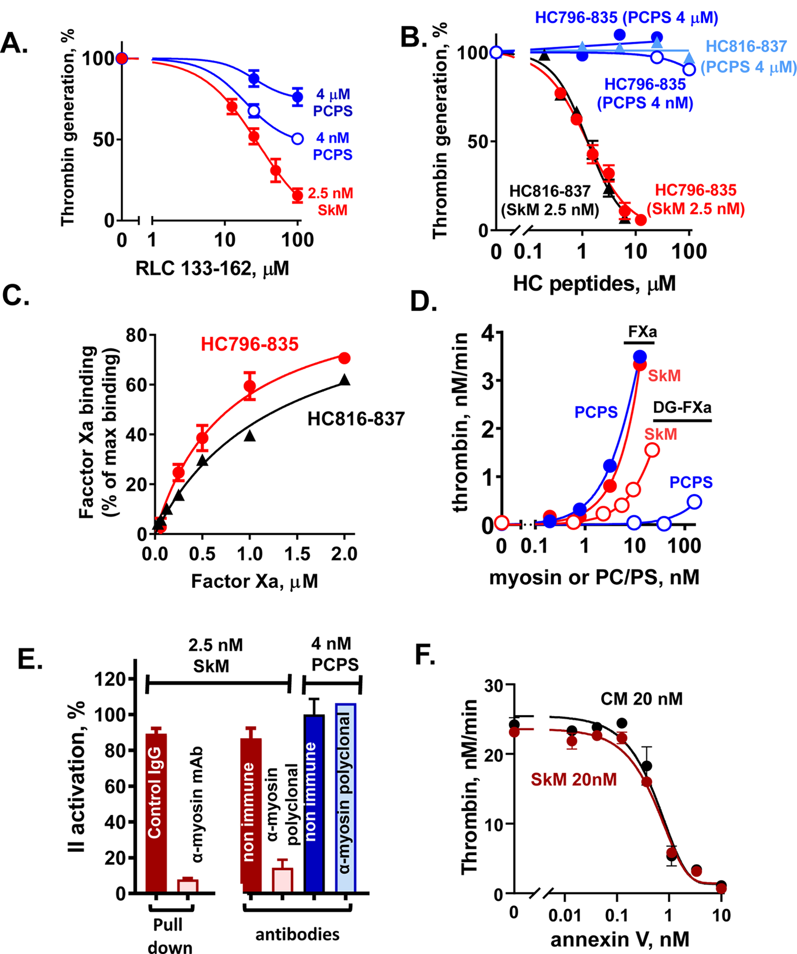
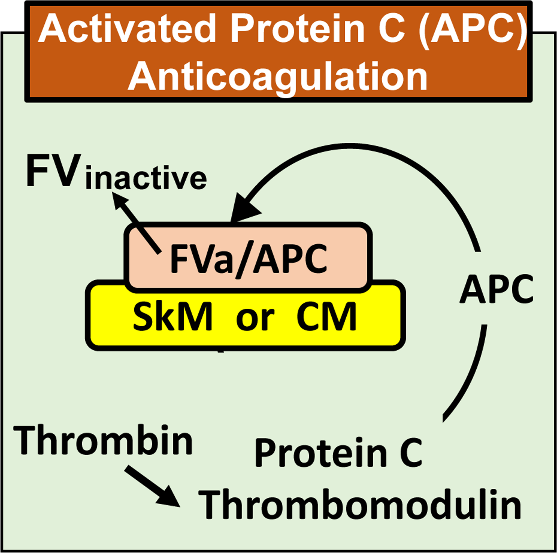
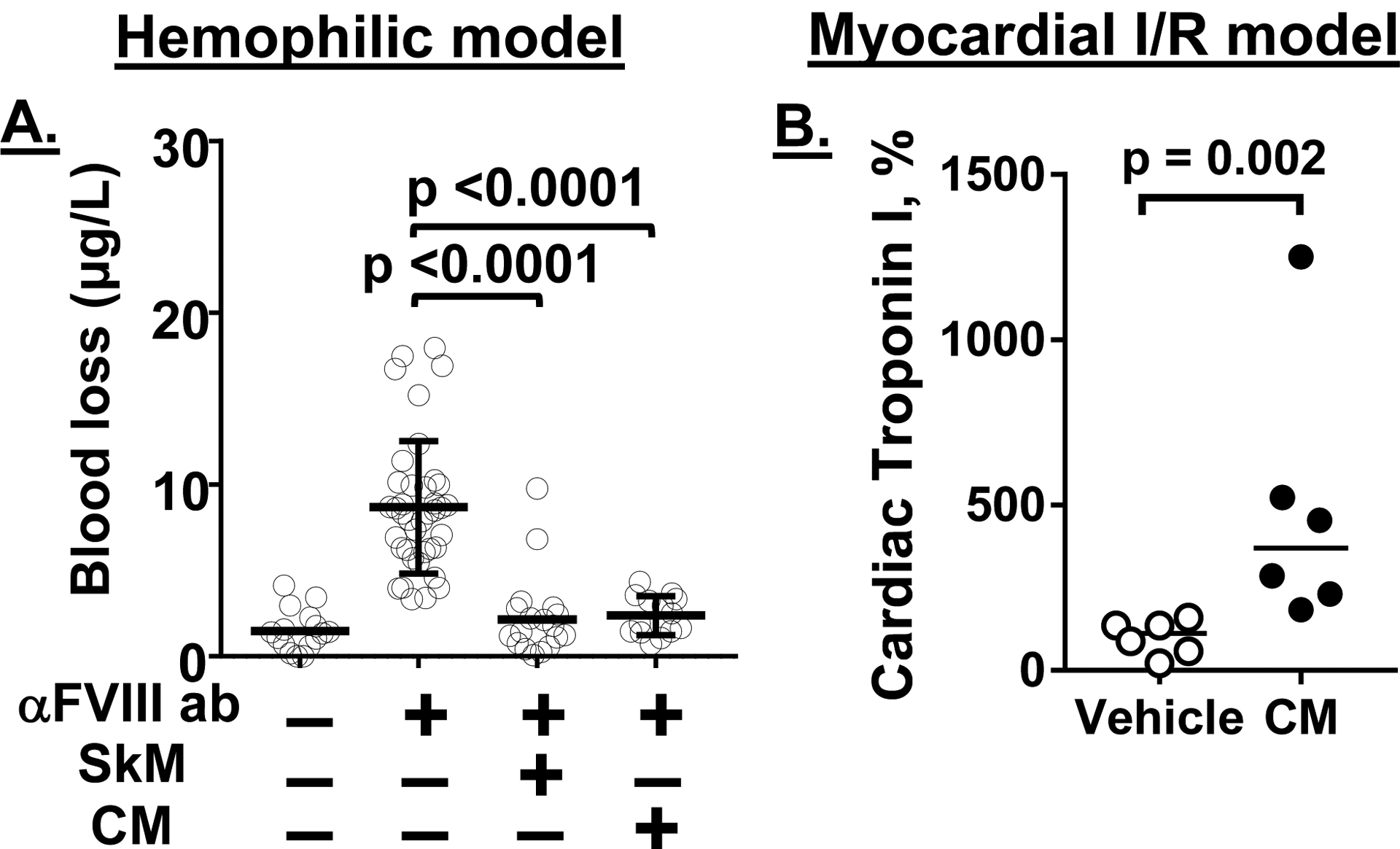
Essentials Striated muscle myosins can promote prothrombin activation by FXa or FVa inactivation by APC. Cardiac myosin and skeletal muscle myosin are pro-hemostatic in murine tail cut bleeding models. Infused cardiac myosin exacerbates myocardial injury caused by myocardial ischemia reperfusion. Skeletal muscle myosin isoforms that circulate in human plasma can be grouped into 3 phenotypes. ABSTRACT: Two striated muscle myosins, namely skeletal muscle myosin (SkM) and cardiac myosin (CM), may potentially contribute to physiologic mechanisms for regulation of thrombosis and hemostasis. Thrombin is generated from activation of prothrombin by the prothrombinase (IIase) complex comprising factor Xa, factor Va, and Ca++ ions located on surfaces where these factors are assembled. We discovered that SkM and CM, which are abundant motor proteins in skeletal and cardiac muscles, can provide a surface for thrombin generation by the prothrombinase complex without any apparent requirement for phosphatidylserine or lipids. These myosins can also provide a surface that supports the inactivation of factor Va by activated protein C/protein S, resulting in negative feedback downregulation of thrombin generation. Although the physiologic significance of these reactions remains to be established for humans, substantive insights may be gleaned from murine studies. In mice, exogenously infused SkM and CM can promote hemostasis as they are capable of reducing tail cut bleeding. In a murine myocardial ischemia-reperfusion injury model, exogenously infused CM exacerbates myocardial infarction damage. Studies of human plasmas show that SkM antigen isoforms of different MWs circulate in human plasma, and they can be used to identify three plasma SkM phenotypes. A pilot clinical study showed that one SkM isoform pattern appeared to be linked to isolated pulmonary embolism. These discoveries enable multiple preclinical and clinical studies of SkM and CM, which should provide novel mechanistic insights with potential translational relevance for the roles of CM and SkM in the pathobiology of hemostasis and thrombosis.

Keywords: blood coagulation; factor X; myocardial infarction; myosin; prothrombinase; thrombin.


Conflict of interest statement

CONFLICTS OF INTEREST

None of the authors has a conflict of interest to declare with regard to the content of this review.

Figures

Figure 1.
Figure 1.. Conventional Myosin Structure and Nomenclature.
(A) Structure of conventional myosins which contain 6 polypeptides, 2 heavy chains (HC), and 4 light chains [2 ‘essential’ and 2 ‘regulatory’ light chains (ELC and RLC, respectively)]. Conventional myosin can be split into 1 light meromyosin (LMM) fragment and 1 heavy meromyosin (HMM) fragment; HMM can be further split into two subfragments, designated S1 and S2 subfragments [23]. (B) Structure of conventional myosin S1 domain containing a neck (N-terminal HC, ELC and RLC, the hypothesized procoagulant surface [24]), a converter domain (HC followed by neck), upper and lower 50 K domains that contain actin and ATP binding sites, in the N-terminal domain [–22]. Structure shown in (B) is adopted with modifications from Rayment et al [21]. (C) Either SkM or CM (myosins) which is depicted on a yellow background can bind factors Xa and Va and thereby can enhance prothrombin activation on its surface. Kinetic studies indicate that the potency for SkM and CM preparations to enhance prothrombin activation is generally comparable to that of procoagulant phospholipid vesicles (Table 1). SkM preparations contain very low levels of phosphatidylserine [15], raising the queston of whether myosin-bound phospholipids may contribute to enhance SkM’s or CM’s procoagulant activity.
Figure 2.
Figure 2.. Dimensions for space-filling structures of FXa:FVa complex and of SkM’s neck region.
(A) 3-dimensional structure computer model for FXa/FVa complex (from Autin et al [27]). (B) Candidate SkM sequences in neck region that are hypothesized to form at least part of the IIase surface in space-filling format. Each structure is displayed as front and back views, related by a 180 degree rotation. (C) Simplified scheme and its length depicting the FXa/FVa interaction sites on the neck region of SkM.
Figure 3.
Figure 3.. Experimental data from studies indicating that SkM and CM proteins are essential for enhancing prothrombin activation.
(A, B) Inhibition of myosin-enhanced or phospholipid vesicle-enhanced prothrombin activation by SkM-sequence based peptides (RLC133–162 (A) and HC796–835 and HC816–836 (B)) was studied in the presence of SkM (2.5 nM, final) or phospholipid vesicles (PC/PS, 80%/20% w/w) (4 μM or 4 nM, final) as described [24]. Thrombin generation was determined as described [24]. 100 % was the value thrombin generation for controls in the absence of added peptides. (C) The binding of FXa, as determined using FXa chromogenic activity assays, to immobilized HC peptides was tested as described [24]. Each value represents the mean [SD] of at least triplicate determinations. (D) The effect of varying concentrations of SkM or of PC/PS (80%/20%) vesicles on the initial rate of prothrombin activation by FXa or Gla domain-less FXa (DG-FXa) (0.2 nM, final) was determined as described [14]. (E) Anti-myosin antibodies were used to pull-down SkM from solution or to block myosin’s procoagulant activity, as described [14]. From left to right, 2 studies are depicted. First, immobilized mAb-MF20 removed > 90 % of SkM’s procoagulant activity from solution vs. control non-immune IgG. Second, polyclonal anti-myosin antibodies in solution blocked > 80% of SkM’s procoagulant activity vs. controls (two red bars), and there were no significant effects on the procoagulant activity of PC/PS vesicles by anti-myosin or non-immune IgG’s (two blue bars). (F) The effects of varying concentrations of annexin V on the initial rate of prothrombin activation by FXa/FVa in the presence of SkM (closed symbols) or CM (open symbols) (each at 20 nM final) were determined. Annexin V was incubated with FVa (2.4 nM, final) and FXa (0.2 nM, final) in Tris-buffered saline, 0.5% BSA plus 5 mM CaCl2 with SkM or CM, and thrombin generation was initiated by adding prothrombin (0.75 μM, final). After 5 min, quenching was made with EDTA (10 mM, final) and thrombin formation was quantified. For all studies described in this figure, as for our published studies [–16, 24, 25], the SkM or CM preparation was dissolved in water and then immediately dialyzed into Tris buffer (pH 7.4) containing 0.6 M NaCl at 4°C. After dialysis, particles causing turbidity were removed by high speed centrifugation (21,130xg for 1 min) which removed visible aggregates.
Figure 4.
Figure 4.
Scheme depicting the ability of myosin to bind FVa and enhance proteolytic inactivation of FVa by activated protein C (APC).
Figure 5.
Figure 5.. Myosin effects on in vivo hemostasis and thrombosis in murine injury models
(A) Tail bleeding in acquired hemophilia A mice. wt-C57BL/6J mice were treated with anti-FVIII Mab to induce a bleeding tendency due to acquired hemophilia A. When SkM or CM (5.4 mg/kg) or vehicle was given i.v. 15 min before tail clipping and then tail bleeding after cutting was quantified, SkM and CM each very significantly reduced blood loss [13, 16]. (B) Murine myocardial ischemia reperfusion injury. When wt-C57BL/6J mice (n=6/group) subjected to myocardial ischemia reperfusion injury were given CM (5.4 mg/kg) or vehicle via intraarterial infusion at 15 min after initiation of reperfusion, serum cardiac troponin I levels measured at 3 hours after injury initiation were increased [16]. 100% was defined as the median of control vehicle group values. Bars show medians and p values were calculated by Mann-Whitney test.

References

    1. Monroe DM, Hoffman M. What does it take to make the perfect clot? Arterioscler Thromb Vasc Biol. 2006; 26: 41–8. 10.1161/01.Atv.0000193624.28251.83. - DOI - PubMed
    1. Furie B, Furie BC. Mechanisms of thrombus formation. N Engl J Med. 2008; 359: 938–49. 10.1056/NEJMra0801082. - DOI - PubMed
    1. Flaumenhaft R. Thrombus formation reimagined. Blood. 2014; 124: 1697–8. 10.1182/blood-2014-06-579656. - DOI - PMC - PubMed
    1. Byrnes JR, Wolberg AS. Red blood cells in thrombosis. Blood. 2017; 130: 1795–9. 10.1182/blood-2017-03-745349. - DOI - PMC - PubMed
    1. Weisel JW, Litvinov RI. Red blood cells: the forgotten player in hemostasis and thrombosis. J. Thromb. Haemost 2019; 17: 271–82. 10.1111/jth.14360. - DOI - PMC - PubMed

Publication types