. 2020 Dec;18(12):3359-3370.
doi: 10.1111/jth.15095. Epub 2020 Oct 5.

Endothelial JAK2V617F mutation leads to thrombosis, vasculopathy, and cardiomyopathy in a murine model of myeloproliferative neoplasm

Affiliations

Endothelial JAK2V617F mutation leads to thrombosis, vasculopathy, and cardiomyopathy in a murine model of myeloproliferative neoplasm

Melissa Castiglione et al. J Thromb Haemost. 2020 Dec.

Abstract

Objective: Cardiovascular complications are the leading cause of morbidity and mortality in patients with myeloproliferative neoplasms (MPNs). The acquired kinase mutation JAK2V617F plays a central role in these disorders. Mechanisms responsible for cardiovascular dysfunction in MPNs are not fully understood, limiting the effectiveness of current treatment. Vascular endothelial cells (ECs) carrying the JAK2V617F mutation can be detected in patients with MPNs. The goal of this study was to test the hypothesis that the JAK2V617F mutation alters endothelial function to promote cardiovascular complications in patients with MPNs.

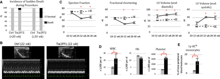
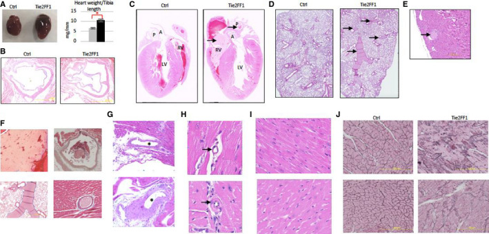
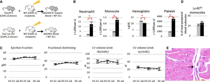
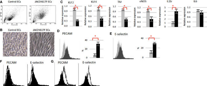
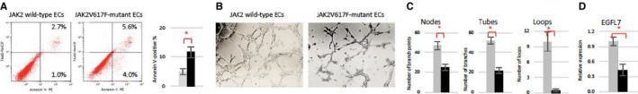
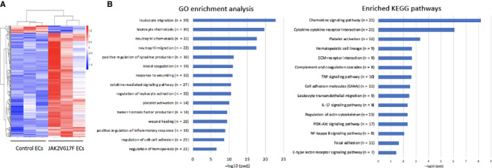
Approach and results: We employed murine models of MPN in which the JAK2V617F mutation is expressed in specific cell lineages. When JAK2V617F is expressed in both blood cells and vascular ECs, the mice developed MPN and spontaneous, age-related dilated cardiomyopathy with an increased risk of sudden death as well as a prothrombotic and vasculopathy phenotype on histology evaluation. In contrast, despite having significantly higher leukocyte and platelet counts than controls, mice with JAK2V617F-mutant blood cells alone did not demonstrate any cardiac dysfunction, suggesting that JAK2V617F-mutant ECs are required for this cardiovascular disease phenotype. Furthermore, we demonstrated that the JAK2V617F mutation promotes a pro-adhesive, pro-inflammatory, and vasculopathy EC phenotype, and mutant ECs respond to flow shear differently than wild-type ECs.

Conclusions: These findings suggest that the JAK2V617F mutation can alter vascular endothelial function to promote cardiovascular complications in MPNs. Therefore, targeting the MPN vasculature represents a promising new therapeutic strategy for patients with MPNs.

Keywords: cardiomyopathy; endothelial cells; myeloproliferative disorders; thrombosis; vascular diseases.


Conflict of interest statement

The authors have declared that no conflicts of interest exists.

Figures

FIGURE 1
FIGURE 1
Development of spontaneous congestive heart failure in Tie2FF1 mice. A, Increased incidence of sudden death in Tie2FF1 mice after 20 weeks of age (n = 14 in each group). B and C, Representative left ventricular echocardiographic tracings (B) and measurement of ejection fraction, fractional shortening, and left ventricular (LV) volume in Tie2 ctrl (dotted line) and Tie2FF1 (black line) mice (C). Results are expressed as mean value ± standard error of the mean. Statistical significance was determined by the Mann‐Whitney test (n = 4 mice in each group at 10–12 weeks and 20–22 weeks, n = 10 mice in each group at 28–30 weeks). D, Blood counts of 20‐ to 22‐week‐old Tie2‐cre control (gray) and Tie2FF1 (black) mice (n = 5–6 in each group). E, Ly‐6Chi (CD11bhiLy‐6GloLy‐6Chi) monocytes in 20‐ to 30‐week‐old Tie2‐cre control (gray) and Tie2FF (black) mice (n = 4 in each group). Statistical significance for (D) and (E) was determined by the Mann‐Whitney test. *P < .05
FIGURE 2
FIGURE 2
The JAK2V617F‐positive Tie2FF1 mice have a prothrombotic and vasculopathy phenotype. A, Representative image of heart (left) and heart weight adjusted by tibia length (right) of 20‐ to 30‐week‐old Tie2 ctrl (gray) and Tie2FF1 (black) mice (n = 4–5 in each group). Statistical significance was determined by the Mann‐Whitney test. B, Representative hematoxylin/eosin (H&E)‐stained aortic root area of Tie2‐Cre ctrl and Tie2FF1 mice (magnification 4×). No significant atherosclerotic lesion was detected. C, Representative H&E staining of longitudinal sections of Tie2‐cre control and Tie2FF1 mice. Note the presence of thrombus (arrow) in right ventricle and main pulmonary artery of the Tie2FF1 mice. LV: left ventricle; RV: right ventricle; A: aortic root; P: main pulmonary artery (magnification 10×). D, Representative H&E staining of lung sections from Tie2‐cre control and Tie2FF1 mice. Note the presence of thrombus (arrow) in segment pulmonary arteries of the Tie2FF1 mice (magnification 10×). E, Representative H&E staining of coronary arteriole thrombus from the Tie2FF1 mice (magnification 10×). F, Representative Masson's trichrome staining demonstrates typical fibrin clot in right ventricle (upper left, 4×), pulmonary artery (upper right, 4×), segment pulmonary arteries (bottom left, 4×), and coronary arteriole (bottom right, 40×) of Tie2FF1 mice. G, Representative H&E staining of epicardial coronary arteries (stars) in Tie2‐cre control (top) and Tie2FF1 (bottom) mice. Note significant vascular wall thickening of the epicardial coronary artery in Tie2FF1 mice (magnification 4×). H, Representative H&E staining of intramyocardial coronary arterioles (arrow) in Tie2‐cre control (top) and Tie2FF1 (bottom) mice. Note stenotic lumen narrowing of the arteriole in Tie2FF1 mice (magnification 40×). I, Representative H&E‐stained cardiac sections taken from similar locations of the heart showing enlarged cardiomyocytes in Tie2FF1 mice (bottom) compared to control mice (top) (magnification 40×). J, Representative reticulin staining of cardiac sections taken from similar locations of the heart from control (left) and Tie2FF1 (right) mice are shown (magnification 20×). (For C–I: 5 control mice and 8 Tie2FF1 experiment mice were examined with similar findings; for J: n = 3 mice in each group were examined.) *P < .05
FIGURE 3
FIGURE 3
Normal heart function in mice with JAK2V617F‐mutant blood cells and wild‐type vascular endothelial cells. A, Experimental scheme to generate a chimeric murine model with JAK2V617F‐mutant blood cells and wild‐type vascular endothelium. B, Blood counts in recipient mice of either Tie2‐cre control (gray) or Tie2FF1 (black) marrow cells 22 weeks after transplantation (n = 5 mice in each group). C, Serial measurements of ejection fraction, fractional shortening, and left ventricular (LV) volumes in recipients of Tie2‐cre control marrow (dotted line) and recipients of Tie2FF1 marrow (black line; n = 5 mice in each group). D, Ly‐6Chi monocytes in recipient mice of Tie2‐Cre control (gray) or Tie2FF1 (black) marrow cells 22 weeks after transplantation (n = 5 mice in each group). E, Representative hematoxylin/eosin staining of intramyocardial coronary arterioles (arrow) in a recipient mouse of Tie2FF1 marrow (magnification 40×). Statistical significance for B–D was determined by the Mann‐Whitney test. *P < .05
FIGURE 4
FIGURE 4
JAK2V617F‐mutant quantitative polymerase chain reaction (ECs) display a pro‐adhesive and pro‐inflammatory phenotype. A, Representative flow cytometry dot plots of wild‐type and JAK2V617F‐mutant ECs. Note JAK2V617F ECs have higher forward scatter (ie, increased cell size) and side scatter (ie, increased granularity) compared to wild‐type (WT) ECs. B, Representative bright field images of wild‐type and JAK2V617F ECs (magnification: 10×). C, Gene expression levels in wild‐type (gray) and JAK2V617F (black) murine ECs were measured using real‐time quantitative polymerase chain reaction. Gene expression in VF ECs is shown as the fold change compared to WT EC expression, which was set as “1.” D and E, Representative flow cytometry histogram plots and quantitative analysis of platelet endothelial cell adhesion molecule (PECAM; D) and E‐selectin (E) expression in un‐sheared wild‐type (gray) and JAK2V617F‐mutant (black) ECs. F and G, Representative flow cytometry histogram plots of PECAM and E‐selectin expression in unsheared (black) vs sheared (white) wild‐type ECs (F) and JAK2V617F ECs (G). *P < .05
FIGURE 5
FIGURE 5
JAK2V617F‐mutant cardiac endothelial cells (ECs) display a vasculopathy phenotype. A, Representative flow cytometry plots (left) and quantitative analysis (right) showing increased apoptosis in cultured JAK2V617F ECs (black bar) compared to wild‐type ECs (gray bar). B, Wild‐type and JAK2V617F‐mutant cardiac ECs (8 × 104) were seeded in Matrigel matrix in 48‐well plate. Representative tube formation after a 4‐hour incubation is shown. Magnification: 10×. C, Quantification of tube formation was performed on images taken at 4× magnification by counting the number of nodes (or branch points), tubes, and loops in four non‐overlapping fields. Results are expressed as the mean ± standard error of the mean (n = 4). Data are from one of two independent experiments on two different pairs of cardiac ECs that gave similar results. D, Gene expression level of EGFL7 in wild‐type (gray) and JAK2V617F (black) cardiac ECs measured using real‐time quantitative polymerase chain reaction. Gene expression in VF ECs is shown as the fold change compared to the average wild‐type EC expression which was set as “1.” (n = 3 in each group) *P < .05
FIGURE 6
FIGURE 6
Transcriptomic profiling of wild‐type and JAK2V617F‐mutant cardiac endothelial cells (ECs). A, Unsupervised hierarchical cluster heatmap of 234 differentially expressed genes between wild‐type and JAK2V617F ECs with an adjusted P‐value < 0.05. B, Differentially enriched Gene Ontology (GO) terms and Kyoto Encyclopedia of Genes and Genomes (KEGG) pathways in JAK2V617F‐mutant cardiac ECs compared to wild‐type control ECs. P values are plotted as the negative of their logarithm

References

    1. Landolfi R, Di Gennaro L, Falanga A. Thrombosis in myeloproliferative disorders: pathogenetic facts and speculation. Leukemia. 2008;22:2020‐2028. - PubMed
    1. Zhan H, Kaushansky K. Functional interdependence of hematopoietic stem cells and their niche in oncogene promotion of myeloproliferative neoplasms: the 159th biomedical version of "it takes two to tango". Exp Hematol. 2019;70:24‐30. - PMC - PubMed
    1. Xie M, Lu C, Wang J, et al. Age‐related mutations associated with clonal hematopoietic expansion and malignancies. Nat Med. 2014;20:1472‐1478. - PMC - PubMed
    1. Genovese G, Kahler AK, Handsaker RE, et al. Clonal hematopoiesis and blood‐cancer risk inferred from blood DNA sequence. N Engl J Med. 2014;371:2477‐2487. - PMC - PubMed
    1. Jaiswal S, Fontanillas P, Flannick J, et al. Age‐related clonal hematopoiesis associated with adverse outcomes. N Engl J Med. 2014;371:2488‐2498. - PMC - PubMed

Publication types