. 2021 Feb;15(2):523-542.
doi: 10.1002/1878-0261.12800. Epub 2020 Dec 31.

Downregulation of miR-326 and its host gene β-arrestin1 induces pro-survival activity of E2F1 and promotes medulloblastoma growth

Affiliations

Downregulation of miR-326 and its host gene β-arrestin1 induces pro-survival activity of E2F1 and promotes medulloblastoma growth

Evelina Miele et al. Mol Oncol. 2021 Feb.

Abstract

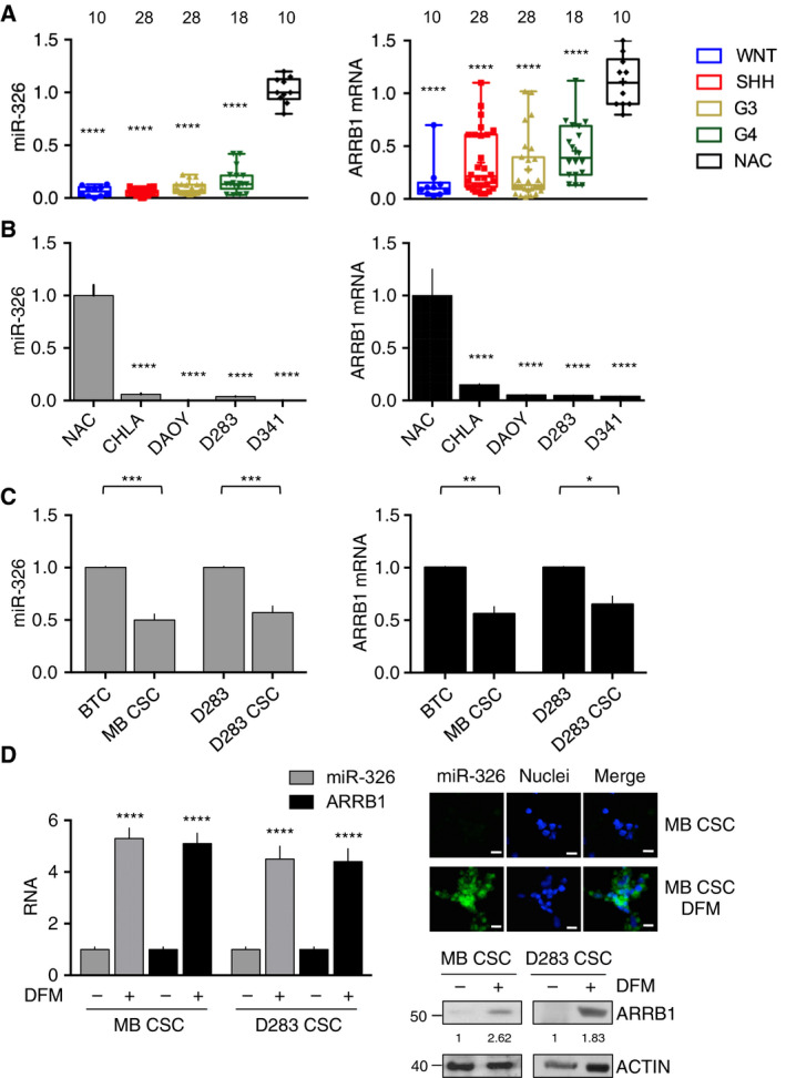
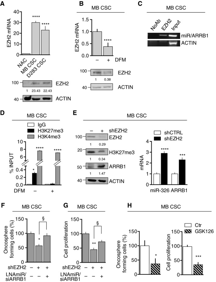
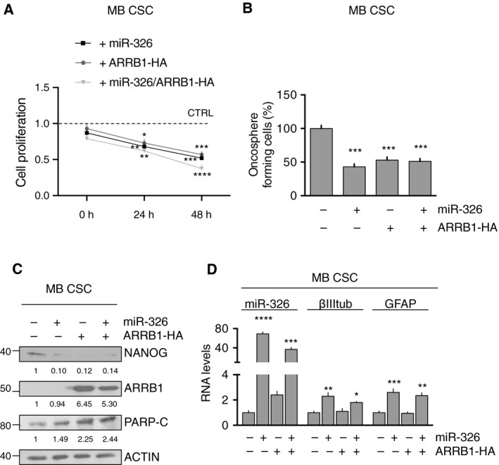
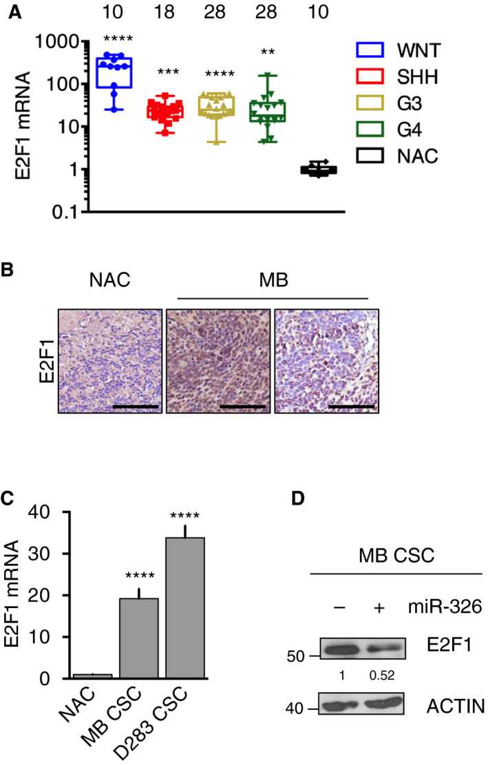
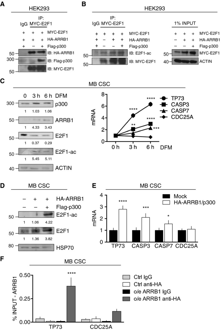
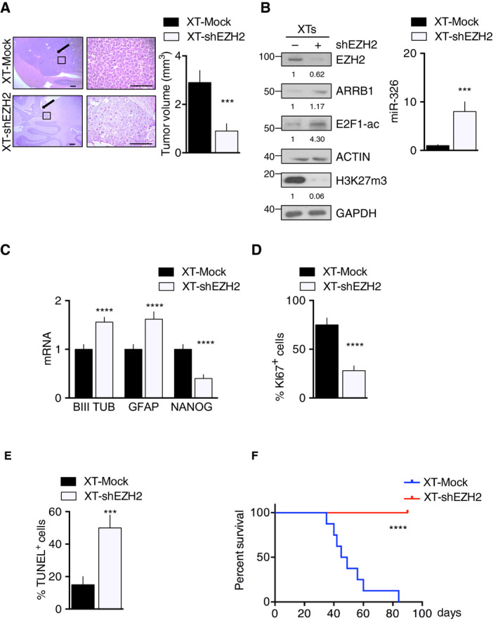
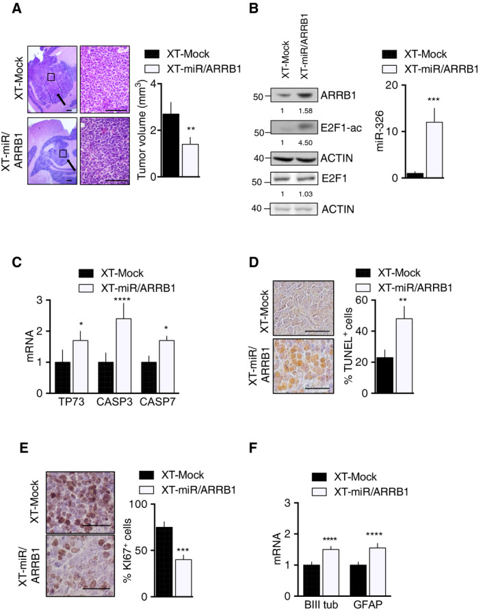
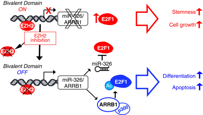
Persistent mortality rates of medulloblastoma (MB) and severe side effects of the current therapies require the definition of the molecular mechanisms that contribute to tumor progression. Using cultured MB cancer stem cells and xenograft tumors generated in mice, we show that low expression of miR-326 and its host gene β-arrestin1 (ARRB1) promotes tumor growth enhancing the E2F1 pro-survival function. Our models revealed that miR-326 and ARRB1 are controlled by a bivalent domain, since the H3K27me3 repressive mark is found at their regulatory region together with the activation-associated H3K4me3 mark. High levels of EZH2, a feature of MB, are responsible for the presence of H3K27me3. Ectopic expression of miR-326 and ARRB1 provides hints into how their low levels regulate E2F1 activity. MiR-326 targets E2F1 mRNA, thereby reducing its protein levels; ARRB1, triggering E2F1 acetylation, reverses its function into pro-apoptotic activity. Similar to miR-326 and ARRB1 overexpression, we also show that EZH2 inhibition restores miR-326/ARRB1 expression, limiting E2F1 pro-proliferative activity. Our results reveal a new regulatory molecular axis critical for MB progression.

Keywords: ARRB1; E2F1; EZH2; medulloblastoma; miR-326.


Conflict of interest statement

The authors declare no conflict of interest.

Figures

Fig. 1
Fig. 1
miR‐326 and ARRB1 under‐expression in MBs and MB cells. (A) qRT‐PCR showed markedly reduced miR‐326 and ARRB1 expression in the 84 MBs of cohort 1 vs. normal adult cerebellum (NAC, control) (miR‐326: WNT/SHH/G3/G4 vs. NAC P < 0.0001; ARRB1: WNT/SHH/G3/G4 vs. NAC P < 0.0001). Numbers of samples tested are indicated above columns. (B) qRT‐PCR revealed miR‐326 and ARRB1 mRNA levels in four MB cell lines. These levels were significantly lower than those in NAC (control) (miR‐326: CHLA, DAOY, D283, D341 vs. NAC P < 0.0001; ARRB1: CHLA, DAOY, D283, D341 vs. NAC P < 0.0001). (C) miR‐326 and ARRB1 transcript levels in CSCs derived from primary MBs (cohort 1, MB CSC1–6) and D283 cells (D283 CSCs) (see Fig. S2) and those found in their respective bulk tumor cell (BTC) populations (miR‐326: MB CSC1–6 vs. BTC1–6 P = 0.0002; D283 CSC vs. D283 P = 0.0005; ARRB1: MB CSC1–6 vs. BTC1–6 P = 0.0025; D283 CSC vs. D283 P = 0.0105). (D) Mean miR‐326 and ARRB1 expression in MB CSCs and D283 CSCs before and after differentiation triggered by transfer from stem cell to differentiation medium (DFM). Left panel: miR‐326 and ARRB1 expression assessed by qRT‐PCR (n = 7) (MB CSC: miR‐326 and ARRB1: DFM+ vs. DFM− P < 0.0001; D283 CSC: miR‐326 and ARRB1: DFM+ vs. DFM− P < 0.0001). Center panel: fluorescence in situ hybridization assessment of miR‐326 (representative images, scale bar, 5 µm); Right panel: ARRB1 expression assessed by western blotting. Error bars represent standard deviation from the means. Statistics: One‐way ANOVA and two‐way ANOVA, *P < 0.05; **P < 0.01; ***P < 0.001; ****P < 0.0001.
Fig. 2
Fig. 2
EZH2‐dependent regulation of miR‐326 and ARRB1. (A) EZH2 expression mRNA (top) and protein (bottom) in NAC (control), MB CSC and D283 CSC (vs. NAC: MB CSC P < 0.0001; D283 CSC P < 0.0001). (B) EZH2 expression (mRNA and protein) in MB CSCs (MB CSC1–3) before and after transfer to DFM. ACTIN: loading control (DFM+ vs. DFM−: P < 0.0001). (C) ChIP of MB CSC (MB CSC1–3) lysates precipitated with anti‐EZH2 antibody or (control) no antibody (NoAb). Eluted DNA was amplified by PCR using primers specific for the miR‐326/ARRB1 regulatory region. ACTIN (shown) and ARRB1 exon 5 and 11 primers (not shown) were used as endogenous nonenriched control regions. (See Table S2 for primer details.) (D) ChIP performed using anti‐H3K27me3 and anti‐H3K4me3 antibodies showing presence of repressive and activating histone marks on the miR‐326 and ARRB1 regulatory region (MB CSC1–4) (in SCM vs. No: H3K27me3 P = 0.017; H3K4me3 P < 0.0001, in DFM vs. No: HeK4me3: P < 0.0001). (E) MB CSCs were transduced with lentiviruses harboring EZH2‐specific shRNA (shEZH2) or (controls) shScramble (shCTRL). Left: Immunoblots showing EZH2, total H3K27me3, and ARRB1 protein levels. Right: ARRB1 mRNA and miR‐326 levels measured by qRT‐PCR (MB CSC1–4) (miR‐326: shEZH2 vs. shCTRL P < 0.0001; ARRB1: shEZH2 vs. shCTRL P = 0.0007). (F, G) MB CSCs were transduced with shEZH2, with or without ARRB1‐specific siRNA (siARRB1) plus LNAmiR‐326, and assayed for (F) self‐renewal, reflected by oncosphere formation (MB CSC1–3) (vs. shEZH2−LNAmiR/siARRB1−: shEZH2+ P = 0.011; shEZH2+LNAmiR/siARRB1+ P = 0.029) (G) and proliferation [MTT assay (MB CSC1–4), (vs. shEZH2−LNAmiR/siARRB1−: shEZH2+ P = 0.0016; shEZH2+LNAmiR/siARRB1+ P = 0.048)]. (H) Oncosphere formation (GSK126 vs. Ctr P = 0.013) (left) and proliferation MTT assay (GSK126 vs. Ctr P = 0.003) (right) of MB CSCs (MB CSC1–4) treated with GSK126 (5 µm for 48 h). All data represent means ± SD from at least three independent experiments. Statistics: Wilcoxon signed‐rank test for paired data, one‐way ANOVA and two‐way ANOVA, *P < 0.05; **P < 0.01; ***P < 0.001; ****P < 0.0001; §< 0.05 vs. indicated controls.
Fig. 3
Fig. 3
Biological effects of ectopic expression of miR‐326 and ARRB1 in MB CSCs. (A–C) MB CSCs were assayed 48 h after transfection with miR‐326 and ARRB1, individually or combined. Mock‐transfected cells served as controls. Ectopic expression of ARRB1 and/or miR‐326 in these cells (A) reduced proliferation (MTT assay) (24 h: vs. CTRL: miR‐326+ P = 0.0072; ARRB1‐HA+ P = 0.0245; miR‐326+ARRB1‐HA+ P = 0.0015; 48 h: vs. CTRL: miR‐326+ P = 0.001; ARRB1‐HA+ P = 0.0005; miR‐326+ARRB1‐HA+ P < 0.0001), (B) diminished the frequency of oncosphere‐forming cells (vs. miR‐326‐ARRB1‐HA−: miR‐326+ P = 0.001; ARRB1‐HA+ P = 0.0005; miR‐326+ARRB1‐HA+ P = 0.0003), (C) decreased NANOG expression and increased the expression of PARP‐C (miR‐326 levels: miR‐326+ P < 0.0001; miR‐326+ARRB1‐HA+ P = 0.0002 vs. miR‐326‐ARRB1‐HA−). (D) qRT‐PCR revealed significantly increased expression of neuronal and glial differentiation markers (βIII tubulin and GFAP, respectively) only in MB CSCs overexpressing miR‐326 (alone or with ARRB1) (vs. Mock βIII tubulin: miR‐326 P = 0.0027; miR‐326 and ARRB1 P = 0.046, GFAP: miR‐326 P = 0.0002; miR‐326 and ARRB1 P = 0.011). Data represent means ± SD from five independent experiments. Statistics: One‐way ANOVA and two‐way ANOVA, *P < 0.05; **P < 0.01; ***P < 0.001; ****P < 0.0001 vs. indicated controls.
Fig. 4
Fig. 4
E2F1 is overexpressed in MBs and is a direct target of miR‐326. (A) qRT‐PCR analysis of the 84 primary MBs of cohort 1 disclosed significant tumor‐related increases (vs. NAC) in the transcription of the miR‐326 target E2F1 (vs. NAC: WNT P < 0.0001; SHH P = 0.0007; G3 P < 0.0001; G4 P = 0.0024). Numbers on top indicate number of samples tested. (B) IHC staining for E2F1 in sections of NAC and representative cohort 1 MB samples. Magnification 40×, Scale: 100 µm. (C) E2F1 mRNA levels in NAC (control) MB CSC and D283 CSC (vs. NAC: MB CSC P < 0.0001; D283 CSC P < 0.0001). (D) Representative immunoblot showing significantly decreased E2F1 expression in MB CSCs after ectopic miR‐326 expression (vs. Mock, mock‐transfected controls, P < 0.0001). Data represent means ± SD from three independent experiments. Statistics: One‐way ANOVA test and Wilcoxon signed‐rank test for paired data, **P < 0.01; ***P < 0.001; ****P < 0.0001 vs. indicated controls.
Fig. 5
Fig. 5
ARRB1 modulates E2F1 acetylation and functions. (A, B) HEK293 cells were transfected with plasmids expressing MYC‐E2F1, HA‐ARRB1 and/or Flag‐p300, as indicated. Total cell extracts were incubated with anti‐MYC antibody (IP), or IgG (negative control) and immunoprecipitated with protein A‐coupled beads. (A) Immunoblotting (IB) of the MYC‐E2F1 precipitate revealed the co‐presence of HA‐ARRB1 and Flag‐p300. (B) Left: Total extracts of HEK293 cells transfected with the indicated plasmids were immunoprecipitated (IP) with anti‐MYC or IgG and immunoblotted (IB) with anti‐acetylated‐E2F1 (E2F1‐ac) and anti‐MYC. Right panel: 1% of the immunoprecipitated cell lysates (INPUT) was immunoblotted with anti‐MYC and anti‐actin (loading control). (C) Untransfected MB CSCs (MB CSC1–3) were assayed under basal conditions (growth in SCM) (0 h, control) and after 3 or 6 h of growth in DFM: (left) immunoblot analysis of endogenous expression ARRB1, p300, E2F1, and E2F1‐ac levels and (right) qRT‐PCR assessment of E2F1 pro‐apoptotic (TP73) and pro‐proliferative (CDC25A) target gene transcription (vs. 0 h: TP73 3 h P < 0.0001, 6 h P < 0.0001; CASP3 3 h P = 0.0023, 6 h P < 0.0001; CASP7 6 h P = 0.0004). (D) MB CSCs transfected with HA‐ARRB1 or HA‐ARRB1 plus Flag‐p300 were assayed for endogenous expression of E2F1 and E2F1‐ac (immunoblotting). (E) MB CSCs (MB CSC1–4) transfected with HA‐ARRB1 plus Flag‐p300 were assayed for expression of pro‐apoptotic and pro‐proliferative E2F1 target genes (qRT‐PCR) (vs. Mock: TP73 P < 0.0001; CASP3 P = 0.0002; CASP7 P = 0.026). (F) Anti‐HA‐ARRB1 ChIP in MB CSCs (MB CSC1–4) 24 h after HA‐ARRB1 overexpression (o/e). Eluted DNA was qPCR‐amplified using specific primers for the regulatory regions of TP73 and CDC25A (o/e ARRB1‐HA: TP73 P < 0.0001). Data represent means ± SD from at least 3 independent experiments. Statistics: One‐way ANOVA and two‐way ANOVA, *P < 0.05; **P < 0.01; ***P < 0.001; ****P < 0.0001 vs. indicated controls.
Fig. 6
Fig. 6
In vivo reactivation of miR‐326 and ARRB1 expression by EZH2 knockdown in MB CSCs. Xenograft tumors (XTs) generated in immunocompromised mice using D283 CSC transduced with lentiviral shEZH2 (XT‐shEZH2) or shScramble (XT‐Mock). (A) Left: Representative images of H&E‐stained, largest‐diameter XT sections. Arrows indicate tumor masses. Magnification: 40× (left column), 400× (right). Scale bar, 100 µm. Right: Bar graphs showing XT volumes on postimplantation day 28. Bars represent means (SD), P = 0.0005. (B, C) XT‐shEZH2 and XT‐Mock expression of (B) (left) EZH2, ARRB1, E2F1‐ac and H3K27me3 proteins (ACTIN, GAPDH: loading control), (left) miR‐326 P = 0.0004, (C) mRNA for markers of differentiation (neuronal: βIIItub P < 0.0001; astrocytic: GFAP P < 0.0001) and stemness (NANOG P < 0.0001). (D, E) XT‐shEZH2 and XT‐Mock were assayed for (D) cell proliferation (Ki67 IHC) P < 0.0001 and (E) apoptosis (TUNEL assay positive cells) P = 0.0003. Data represent means ± SD from three independent experiments. Statistics: Wilcoxon signed‐rank test for paired data and two‐way ANOVA test, ***P < 0.001, ****P < 0.0001 vs. relative controls. (F) Kaplan–Meier analysis of survival for mice bearing XT‐shEZH2 vs. XT‐Mock (n = 8 per group), P < 0.0001.
Fig. 7
Fig. 7
Ectopic expression of miR‐326 and ARRB1 inhibits MB growth in vivo. Orthotopic XTs were generated in immunocompromised mice by injection of D283 CSCs transduced with separate vectors overexpressing miR‐326 and ARRB1 (XT‐miR/ARRB1) or empty vector (XT‐Mock, controls). (A) Representative images of H&E‐stained XTs (largest‐diameter sections). Arrows indicate tumor masses. Bar graph: Mean XT volumes at animal sacrifice (28 days postimplantation) P = 0.0043. XTs were assayed for (B) ARRB1 and E2F1‐ac protein levels (left), miR‐326 levels (right) P = 0.0001; (C) expression of pro‐apoptotic E2F1 target genes (qRT‐PCR) (TP73 P = 0.0226; CASP3 P < 0.0001; CASP7 P = 0.002); (D) apoptosis (TUNEL assay, P = 0.0018); (E) cell proliferation (Ki67 IHC, P = 0.0001); (F) and expression of neuronal and glial differentiation markers (βIIItub P < 0.0001 and GFAP P < 0.0001, respectively). Magnification in panels A, D, and E: 4×, 40×, 63×; all scale bars, 100 µm. Data represent means ± SD from eight independent experiments. Statistics: Wilcoxon signed‐rank test for paired data and two‐way ANOVA, *P < 0.05; **P < 0.01; ***P < 0.001; ****P < 0.0001 vs. indicated controls.
Fig. 8
Fig. 8
Impact of EZH2—miR‐326/ARRB1—E2F1 axis in MB CSCs. In MB CSCs, the miR‐326 and ARRB1 transcription unit remains in a poised state: ready to be transcribed thanks to the presence of the permissive (H3K4me3) chromatin mark, but prevented from doing so by the persistence/predominance of the repressive (H3K27me3) chromatin mark, which is catalyzed by the histone methyltransferase EZH2. In this state, the cells express high levels of nonacetylated E2F1 transcription factor, which favors their self‐renewal and proliferation. Reversal of the H3K4me3: H3K27me3 ratio de‐represses miR‐326 and ARRB1 transcription. Restoration of miR‐326 expression reduces the E2F1 levels. Re‐expression of ARRB1, in complex with p300, acetylates E2F1 (E2F1‐Ac), thereby redirecting the transcription factor's activity toward pro‐apoptotic gene targets (e.g., TP73, CASP3, CASP7).

Similar articles

Cited by

References

    1. Kool M, Korshunov A, Remke M, Jones D, Schlanstein M, Northcott P, Cho Y‐J, Koster J, Schouten‐van Meeteren A, van Vuurden D et al (2012) Molecular subgroups of medulloblastoma: an international meta‐analysis of transcriptome, genetic aberrations, and clinical data of WNT, SHH, Group 3, and Group 4 medulloblastomas. Acta Neuropathol 123, 473–484. - PMC - PubMed
    1. Hovestadt V, Remke M, Kool M, Pietsch T, Northcott PA, Fischer R, Cavalli FMG, Ramaswamy V, Zapatka M, Reifenberger G et al (2013) Robust molecular subgrouping and copy‐number profiling of medulloblastoma from small amounts of archival tumour material using high‐density DNA methylation arrays. Acta Neuropathol 125, 913. - PMC - PubMed
    1. Northcott PA, Buchhalter I, Morrissy AS, Hovestadt V, Weischenfeldt J, Ehrenberger T, Gröbner S, Segura‐Wang M, Zichner T, Rudneva VA et al (2017) The whole‐genome landscape of medulloblastoma subtypes. Nature 547, 311–317. - PMC - PubMed
    1. Cavalli FM, Remke M, Rampasek L, Peacock J, Shih DJ, Luu B, Garzia L, Torchia J, Nor C, Morrissy AS et al (2017) Intertumoral heterogeneity within medulloblastoma subgroups. Cancer Cell 31, 737–754, e6. - PMC - PubMed
    1. Northcott PA, Robinson GW, Kratz CP, Mabbott DJ, Pomeroy SL, Clifford SC, Rutkowski S, Ellison DW, Malkin D, Taylor MD et al (2019) Medulloblastoma. Nat Rev Dis Primers 5, 1–20. - PubMed

Publication types

MeSH terms