Review
. 2020 Nov 20;295(47):16121-16155.
doi: 10.1074/jbc.REV120.008029. Epub 2020 Sep 13.

Emerging roles of the MAGE protein family in stress response pathways

Affiliations
Review

Emerging roles of the MAGE protein family in stress response pathways

Rebecca R Florke Gee et al. J Biol Chem. .

Abstract

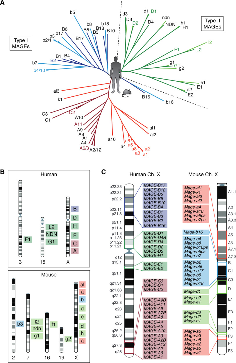
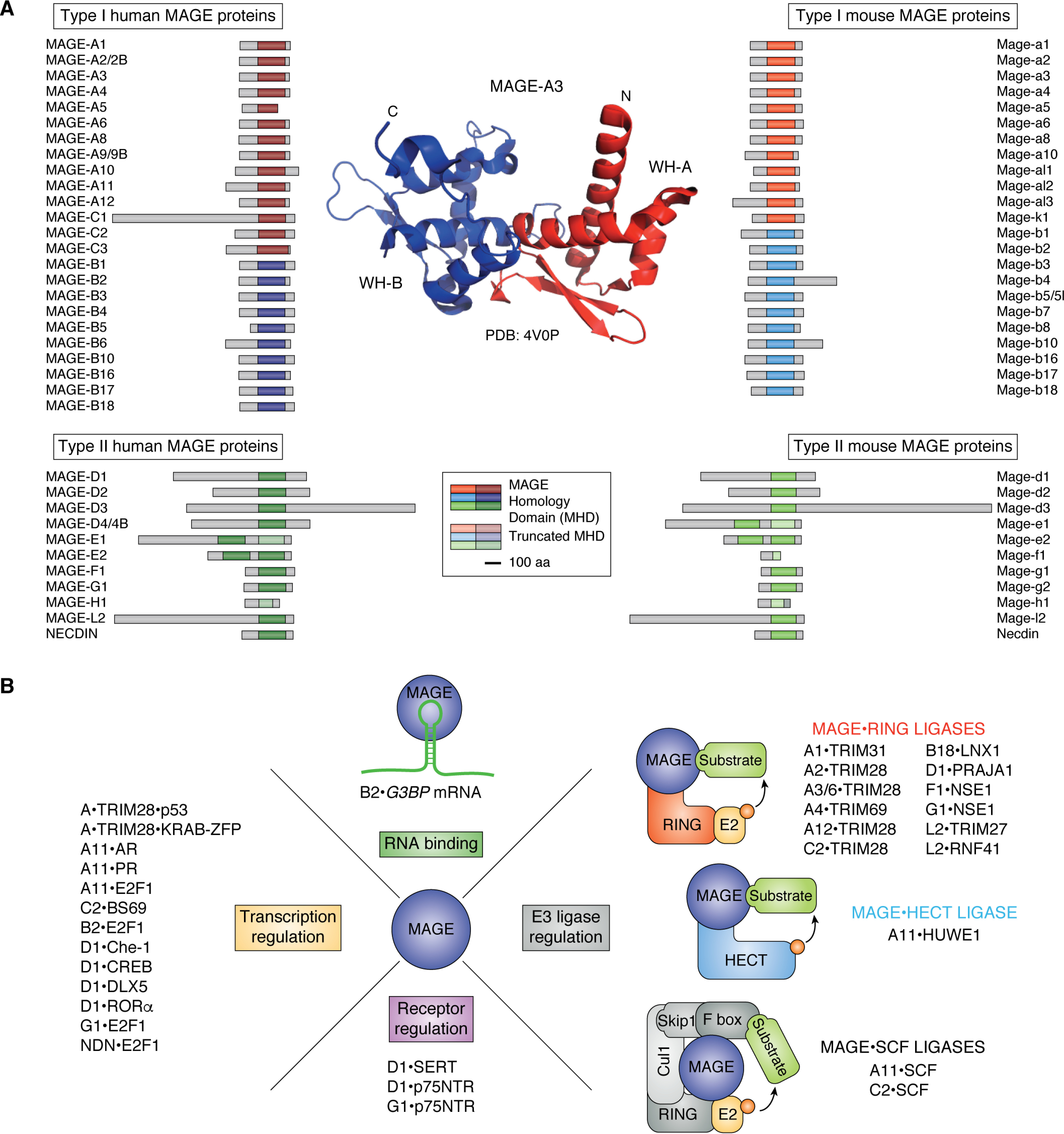
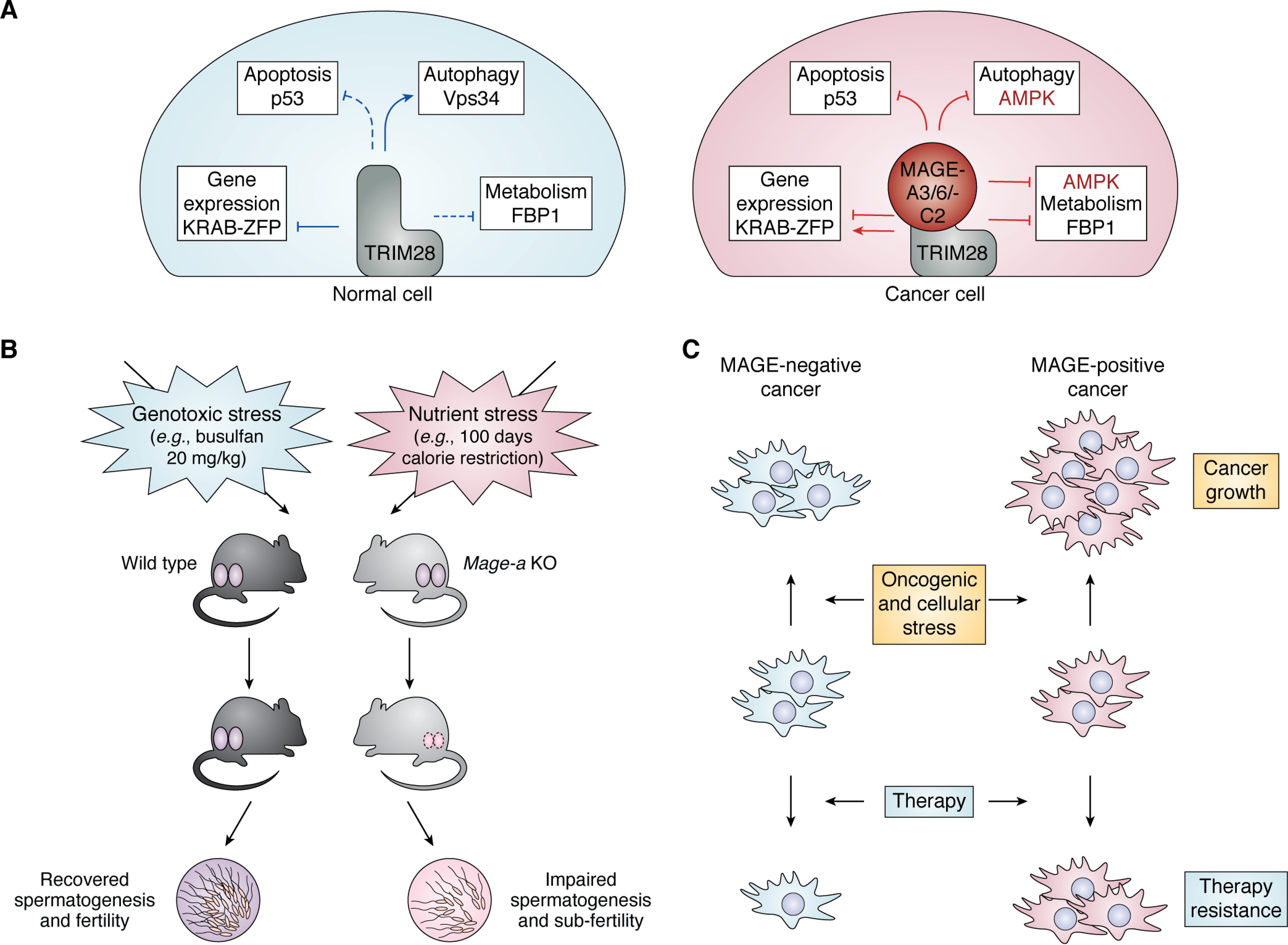
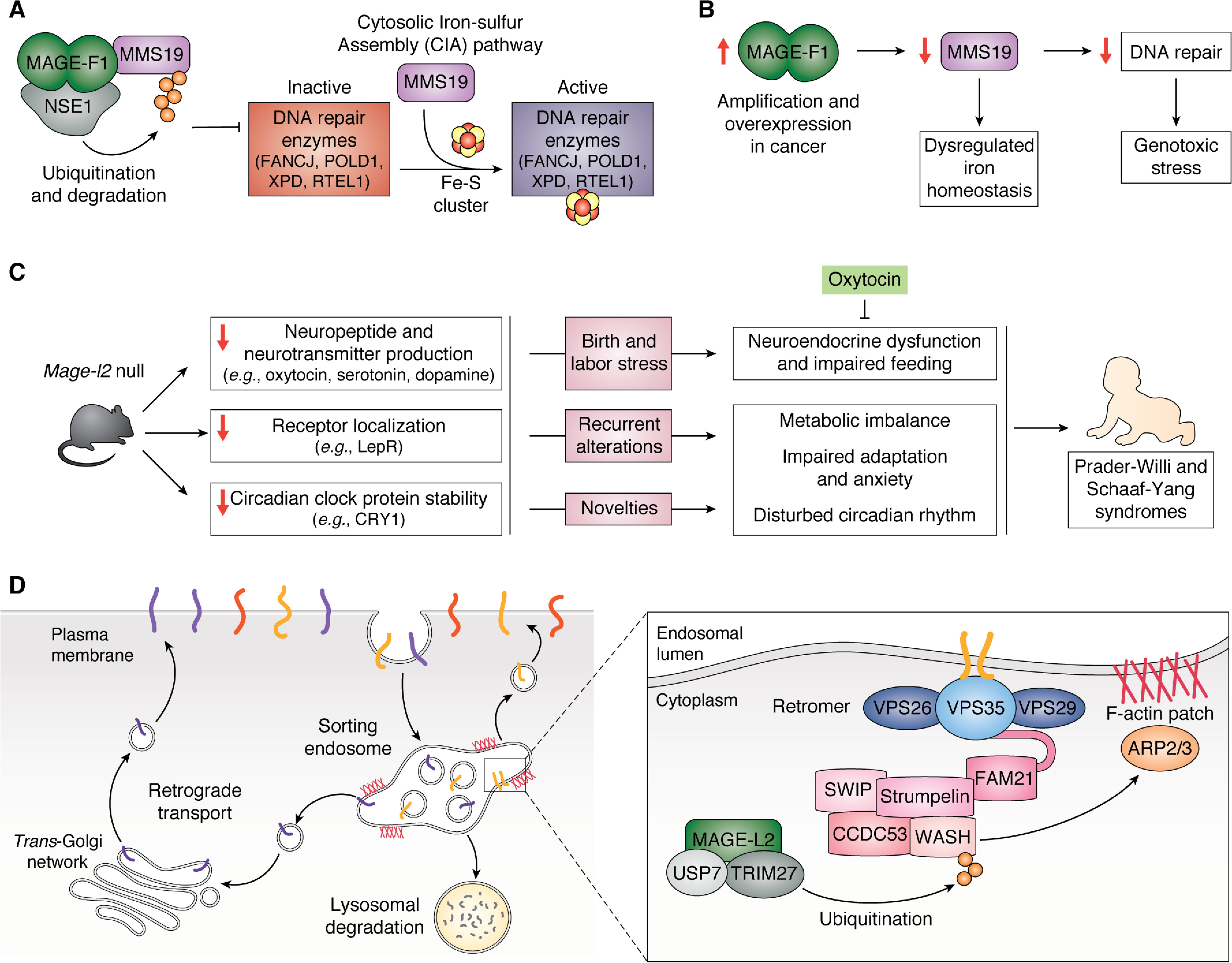
The melanoma antigen (MAGE) proteins all contain a MAGE homology domain. MAGE genes are conserved in all eukaryotes and have expanded from a single gene in lower eukaryotes to ∼40 genes in humans and mice. Whereas some MAGEs are ubiquitously expressed in tissues, others are expressed in only germ cells with aberrant reactivation in multiple cancers. Much of the initial research on MAGEs focused on exploiting their antigenicity and restricted expression pattern to target them with cancer immunotherapy. Beyond their potential clinical application and role in tumorigenesis, recent studies have shown that MAGE proteins regulate diverse cellular and developmental pathways, implicating them in many diseases besides cancer, including lung, renal, and neurodevelopmental disorders. At the molecular level, many MAGEs bind to E3 RING ubiquitin ligases and, thus, regulate their substrate specificity, ligase activity, and subcellular localization. On a broader scale, the MAGE genes likely expanded in eutherian mammals to protect the germline from environmental stress and aid in stress adaptation, and this stress tolerance may explain why many cancers aberrantly express MAGEs Here, we present an updated, comprehensive review on the MAGE family that highlights general characteristics, emphasizes recent comparative studies in mice, and describes the diverse functions exerted by individual MAGEs.

Keywords: AMP-activated kinase (AMPK); DNA damage response; E3 ligase; E3 ubiquitin ligase; Fe-S cluster; MAGE; Prader-Willi syndrome; Schaaf-Yang syndrome; alternative polyadenylation; apoptosis; cancer; cancer-testis antigen; cell metabolism; melanoma antigen; metabolism; p53; spermatogenesis; stress adaptation; stress granule; stress response; ubiquitin; ubiquitination.


Conflict of interest statement

Conflict of interest—P. R. P. is a paid consultant of Levo Therapeutics, Inc. and Amgen, Inc.

Figures

Figure 1.
Figure 1.
Overview of the MAGE gene family in humans and mice. A, phylogenetic tree showing the relationship between human and mouse MAGE proteins. The tree was created by the neighbor-joining construction method using the Jukes–Cantor protein distance measurement from the CLC Main Workbench 20. B, chromosomal location of human and mouse MAGE genes. C, locations of MAGE genes on the human and mouse X chromosome based on the recent NCBI's genome assembly HRCh38.p13 and GRCm38.p6. For all figures, the type II MAGEs are represented in green, MAGE-A and -C subfamilies in red, and MAGE-B subfamily in blue. Light colors indicate mouse Mages and dark colors indicate human MAGEs.
Figure 2.
Figure 2.
Expression of MAGEs in normal tissues and cancer. A, human and mouse MAGE expression during different life stages is indicated. Starting with the top part of the outer circle, the expression of MAGEs is depicted during spermatogenesis, in ES cells, in an embryo, and finally in adults. B, the heatmap displays the percentage of various tumors that express each type I MAGE. The results are based upon data generated by the TCGA Research Network (RRID:SCR_003193).
Figure 3.
Figure 3.
General structure of the MHD and biochemical function of MAGE proteins. A, schematic structure of human and mouse MAGE proteins. The MHD for each MAGE is indicated by a solid colored box, and the size corresponding to 100 amino acids (aa) is shown. The crystal structure of the double winged-helix motif of the MHD of MAGE-A3 (Protein Data Bank entry 4V0P) is shown. The N- and C termini are indicated, and the two WH motifs (WH-A and WH-B) are represented in red and blue, respectively. B, MAGEs bind to and regulate E3 ligases, receptors, transcription factors, and RNA (as an RNA-binding protein) to exert diverse molecular functions (General characteristics of MAGE proteins).
Figure 4.
Figure 4.
MAGE-A3/6 and -C2 are cell type–specific regulators of TRIM28 that confer stress resistance to male germline and cancer. A, MAGE-A3/6 and -C2 act as specific regulators of TRIM28 function in transcriptional regulation, apoptosis, autophagy, and cell metabolism (MAGE-A3/6 and MAGE-C2 are cancer cell-specific regulators of TRIM28). B, after genotoxic or nutritional stress, recovery of spermatogenesis in Mage-a KO mice is compromised compared with WT mice. C, MAGE-A3/6 and -C2 promote cancer growth and enable therapy resistance, likely by protecting cells against diverse stressors they encounter during tumorigenesis and treatment.
Figure 5.
Figure 5.
MAGE-A11 and -B2 affect transcription and translation, respectively. A, by binding to the E3 ligase HUWE1, MAGE-A11 specifies PCF11 for ubiquitination, which displaces CFIm25 from the mRNA 3′-end processing complex. The subsequent remodeling of the complex leads to 3′-UTR shortening, which leads to increased levels of oncogenes through loss of miRNA repression. Additionally, down-regulation of ceRNAs inhibits tumor suppressors. Thus, MAGE-A11 function in APA contributes to tumorigenesis. B, MAGE-B2 binds to the G3BP mRNA transcript to repress its translation and decrease G3BP protein concentration. As a result, MAGE-B2/-b4 inhibit stress granule formation and promote cellular stress tolerance, giving a growth advantage to cancer cells and heat tolerance to male germ cells.
Figure 6.
Figure 6.
Functions and pathways of the type II MAGEs, MAGE-F1 and -L2. A, MAGE-F1 controls flux through the CIA pathway by regulating MMS19 protein levels. MAGE-F1 interacts with the E3 ligase NSE1 to form an MRL that ubiquitinates MMS19, promoting its degradation. Decreased MMS19 protein levels lead to decreased iron-sulfur (Fe-S) cluster incorporation into downstream targets, like DNA repair enzymes. B, increased levels of MAGE-F1 contribute to dysregulated iron homeostasis by degrading MMS19 and genotoxic stress by suppressing DNA repair pathways. C, MAGE-L2 contributes to the neurodevelopmental disorders PWS and SYS. Mage-l2–null mice exhibit decreased levels of mature neuropeptides, transmembrane receptors, like LepR, and circadian rhythm proteins, all of which may contribute to impaired adaptation to recurring and acute changes in the environment and contribute to the phenotypes seen in KO mice and PWS and SYS patients. Administration of oxytocin immediately after birth or during the first postnatal week rescues survival and normal adult social behavior of Mage-l2–null mice. D, MAGE-L2 is involved in endosomal protein trafficking. Cargo proteins on endosomes are trafficked to either the plasma membrane, the trans-Golgi network, or the lysosome for degradation. The retromer complex (blue) recognizes cargo proteins and, based on their destination, sorts them into endosomal tubules reshaped by localized F-actin patches. VPS35 interacts with the WASH regulatory complex (pink). The MUST complex (MAGE-L2-USP7-TRIM27) activates WASH by adding a Lys-63–linked polyubiquitin chain, which recruits ARP2/3 and promotes downstream F-actin nucleation.

References

    1. Dunn G. P., Old L. J., and Schreiber R. D. (2004) The three Es of cancer immunoediting. Annu. Rev. Immunol. 22, 329–360 10.1146/annurev.immunol.22.012703.104803 - DOI - PubMed
    1. Whitehurst A. W. (2014) Cause and consequence of cancer/testis antigen activation in cancer. Annu. Rev. Pharmacol. Toxicol. 54, 251–272 10.1146/annurev-pharmtox-011112-140326 - DOI - PubMed
    1. van der Bruggen P., Traversari C., Chomez P., Lurquin C., De Plaen E., Van den Eynde B., Knuth A., and Boon T. (1991) A gene encoding an antigen recognized by cytolytic T lymphocytes on a human melanoma. Science 254, 1643–1647 10.1126/science.1840703 - DOI - PubMed
    1. Jager E., and Knuth A. (2012) The discovery of cancer/testis antigens by autologous typing with t cell clones and the evolution of cancer vaccines. Cancer Immun. 12, 6 - PMC - PubMed
    1. Chomez P., De Backer O., Bertrand M., De Plaen E., Boon T., and Lucas S. (2001) An overview of the mage gene family with the identification of all human members of the family. Cancer Res. 61, 5544–5551 - PubMed

Publication types

MeSH terms

Associated data