

The advantages of carbon fiber based orthopedic devices in patients who have to undergo radiotherapy
- PMID: 32921754
- PMCID: PMC7716998
- DOI: 10.23750/abm.v91i3.7769
The advantages of carbon fiber based orthopedic devices in patients who have to undergo radiotherapy
Abstract
Background and objectives: The modern approach to primary and secondary muscular skeletal tumors is multidisciplinary. The right combination of chemotherapy, surgery and radiotherapy (RT) makes obtaining local and distant disease control more likely. When surgery is indicated, radiotherapy often has a fundamental role as an adjuvant treatment; however, the titanium alloy instrumentations interfere with Radiotherapy setting, decreasing its effectiveness. It is common opinion that carbon fiber-reinforced devices are convenient in case of adjuvant RT in muscular skeletal oncology. The aim of the study is to support this intuition with experimental data, verifying the more accurate estimation of the delivered dose during RT, comparing Carbon Fiber-Reinforced PEEK (CFRP) plates with titanium-alloy orthopedic devices in order to evaluate their effects on target volume identification and dose distribution for radiation treatment.
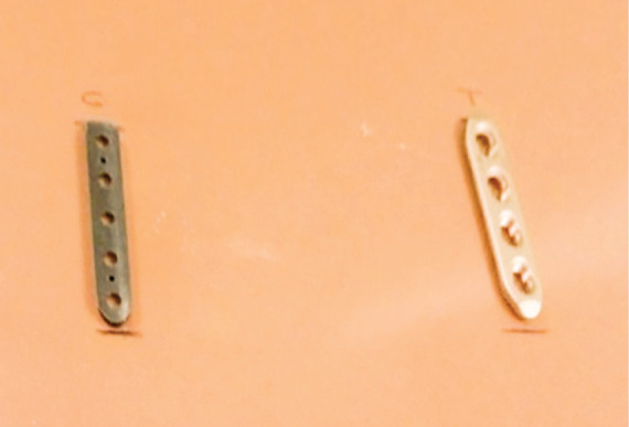
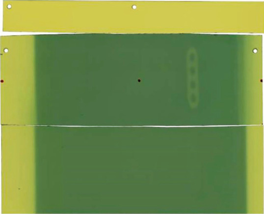
Methods: Phantoms were then irradiated with a linear accelerator Varian 2100 C/D with photon beams of 6 and 15 MV energies. Absorbed dose in the point of interest was verified by EBT3 gafchromic films above and below the two materials. Images from CT simulations were also analyzed in terms of Hounsfield numbers in patients with titanium and carbon fiber orthopedic implants in the spine or in the femur.
Results: For a 6 MV photon beam, the doses measured just under the titanium-alloy plate were less than approximately 20% of the value calculated by the TPS. For a 15 MV beam energy, these differences were slightly lower. Using CFRP plate, the difference between measured and calculated doses was within ±3% for both energies, which was comparable with the statistical uncertainties. In the cases of simulated treatment of humerus titanium implants, the difference varies in range ± 10% with hot spot of + 10% and cold spot of -15%.
Conclusions: The use of CFRP for orthopedic devices and implants provides a valuable advantage in identifying the target due to the reduction of artifacts. Clear imaging of the soft tissues surrounding the bone is useful and reduces the discrepancies between calculated/delivered and measured doses, generating a more homogeneous dose distribution. Furthermore, there is a significant benefit in detecting the state of disease in CT imaging during the follow-up of treated patients. In-vivo studies are encouraged to verify whether a more effective radiotherapy leads to a decrease in local recurrence and local progression.
Conflict of interest statement
Each author declares that he or she has no commercial associations (e.g. consultancies, stock ownership, equity interest, patent/licensing arrangement etc.) that might pose a conflict of interest in connection with the submitted article
No funding was received for publishing the present paper.
Figures


References
-
- Ding GX, Yu CW. A study on beams passing through hip prosthesis for pelvic radiation treatment. Int J Radiat Oncol Biol Phys. 2001;51(4):1167–75. - PubMed
-
- Reft C, Alecu R, Das IJ, et al. Dosimetric considerations for patients with HIP prostheses undergoing pelvic irradiation. Report of the AAPM Radiation Therapy Committee Task Group 63. Med Phys. 2003;30(6):1162–82. - PubMed
-
- Steinberg EL, Rath E, Shlaifer A, et al. : Carbon fiber reinforced PEEK Optima--a composite material biomechanical properties and wear/debris characteristics of CF-PEEK composites for orthopedic trauma implants. J Mech Behav Biomed Mater. 2013;17:221–8. - PubMed
MeSH terms
Substances
LinkOut - more resources
Full Text Sources
