. 2020 Aug 25:15:6451-6468.
doi: 10.2147/IJN.S258906. eCollection 2020.

Tumor Microenvironmental Responsive Liposomes Simultaneously Encapsulating Biological and Chemotherapeutic Drugs for Enhancing Antitumor Efficacy of NSCLC

Affiliations

Tumor Microenvironmental Responsive Liposomes Simultaneously Encapsulating Biological and Chemotherapeutic Drugs for Enhancing Antitumor Efficacy of NSCLC

Liang Kong et al. Int J Nanomedicine. .

Erratum in

Abstract

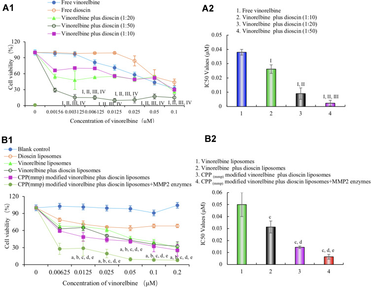
Background: Non-small cell lung cancer (NSCLC) is one of the most lethal types of cancer with highly infiltrating. Chemotherapy is far from satisfactory, vasculogenic mimicry (VM) and angiogenesis results in invasion, migration and relapse.

Purpose: The objective of this study was to construct a novel CPP (mmp) modified vinorelbine and dioscin liposomes by two new functional materials, DSPE-PEG2000-MAL and CPP-PVGLIG-PEG5000, to destroy VM channels, angiogenesis, EMT and inhibit invasion and migration.

Methods and results: The targeting liposomes could be enriched in tumor sites through passive targeting, and the positively charged CPP was exposed and enhanced active targeting via electrostatic adsorption after being hydrolyzed by MMP2 enzymes overexpressed in the tumor microenvironment. We found that CPP (mmp) modified vinorelbine and dioscin liposomes with the ideal physicochemical properties and exhibited enhanced cellular uptake. In vitro and in vivo results showed that CPP (mmp) modified vinorelbine and dioscin liposomes could inhibit migration and invasion of A549 cells, destroy VM channels formation and angiogenesis, and block the EMT process. Pharmacodynamic studies showed that the targeting liposomes had obvious accumulations in tumor sites and magnificent antitumor efficiency.

Conclusion: CPP (mmp) modified vinorelbine plus dioscin liposomes could provide a new strategy for NSCLC.

Keywords: MMP2 enzymes; dioscin; multi-functional liposomes; non-small cell lung cancer; tumor microenvironment; vinorelbine.


Conflict of interest statement

All authors declare that there are no conflicts of interest.

Figures

Figure 1
Figure 1
Schematic illustration of strategy for treating NSCLC by CPP (mmp) modified vinorelbine plus dioscin liposomes. (A) A schematic representation of enzymatic hydrolysis of CPP (mmp) modified vinorelbine plus dioscin liposomes. (B) The enhanced transport drug carrier to tumors and enzymatic hydrolysis to expose CPP. (C) Anti-apoptosis and the inhibition of VM channels formation mediated by vinorelbine and dioscin, respectively.
Figure 2
Figure 2
Characterization of CPP (mmp) modified vinorelbine plus dioscin liposomes. (A) MALDI-TOF-MS spectrum of DSPE-PEG2000-MAL; (B) MALDI-TOF-MS spectrum of DSPE-PEG2000-CPP-PVGLIG-PEG5000; (C) MALDI-TOF-MS spectrum of DSPE-PEG2000-CPP; (D) TEM image of CPP (mmp) modified vinorelbine plus dioscin liposomes, scalar bar=100 nm; (E) AFM image of CPP (mmp) modified vinorelbine plus dioscin liposomes, scale bar=1 μm; (F) the amplified structure of (E), scale bar=500 nm; (G) particle size of CPP (mmp) modified vinorelbine plus dioscin liposomes; (H) particle size of CPP (mmp) modified vinorelbine plus dioscin liposomes incubated with MMP-2 enzymes; (I) Zeta potential of CPP (mmp) modified vinorelbine plus dioscin liposomes; (J) Zeta potential of CPP (mmp) modified vinorelbine plus dioscin liposomes incubated with MMP-2 enzymes.
Figure 3
Figure 3
Cellular uptake and localization after incubation with varying formulations. (A) Cellular uptake of A549 cells treated with the varying liposomal formulations or free vinorelbine; (B) quantitative analysis of fluorescence intensity; 1. Blank control; 2. Epirubicin liposomes; 3. Epirubicin plus dioscin liposomes; 4. CPP (mmp) modified epirubicin plus dioscin liposomes; 5. CPP (mmp) modified epirubicin plus dioscin liposomes + MMP2 enzymes; 6. Free epirubicin; Data are presented as mean ± SD (n=3). I, vs 1; II, vs 2; III, vs 3; IV, vs 4; V, vs 5. P<0.05; (C) cellular uptake of A549 cells treated with CPP (mmp) modified epirubicin plus dioscin liposomes incubated with different concentrations of MMP2 enzymes; (D) quantitative analysis of fluorescence intensity; a. Blank control; b. CPP (mmp) modified epirubicin plus dioscin liposomes without MMP2 enzymes; c. CPP (mmp) modified epirubicin plus dioscin liposomes incubated with MMP2 enzymes (0.2 μM); d. CPP (mmp) modified epirubicin plus dioscin liposomes incubated with MMP2 enzymes (0.5 μM); e. CPP (mmp) modified epirubicin plus dioscin liposomes incubated with MMP2 enzymes (1 μM); Data are presented as mean ± SD (n=3). i, vs a; ii, vs b; iii, vs c; iv, vs d; P<0.05; (E) analysis of localization of A549 cells incubated with the varying formulations by laser scanning confocal microscopy, scale bar=100 μm (n=3). (F) SEM photographs of A549 spheroids. a’. Blank control; b’. Dioscin liposomes; c’. Vinorelbine liposomes; d’. Vinorelbine plus dioscin liposomes; e’. CPP (mmp) modified vinorelbine plus dioscin liposomes; f’. CPP (mmp) modified vinorelbine plus dioscin liposomes incubated with MMP2 enzymes.
Figure 4
Figure 4
Cytotoxic effects on A549 cells after treatments with varying formulations. (A) Cytotoxic effects of free drugs; I, vs Free vinorelbine; II, vs Free dioscin; III, vs Free vinorelbine plus dioscin (1:10); IV, vs Free vinorelbine plus dioscin (1:20); (B) cytotoxic effects of the liposomal formulations. a, vs Blank control liposomes; b, vs Dioscin liposomes; c, vs Vinorelbine liposomes; d, vs Vinorelbine plus dioscin liposomes; e, vs CPP (mmp) modified vinorelbine plus dioscin liposomes. Data are presented as mean±SD (n=6). P<0.05.
Figure 5
Figure 5
Inhibitory effects on invasion, migration and EMT in A549 cells after treatment with varying liposomal formulations. (A) Representative images of A549 cells invasion after treatment with the varying liposomal formulations, scale bar=50 μm; (B) representative images of A549 cells wound closure following treatment with the varying liposomal formulations after 24 h, scale bar=50 μm; (C) representative images of inhibition of A549 cells EMT by the varying liposomal formulations, scale bar=50 μm; (D) semi-quantitative analysis of relative invasion rate of A549 cells after treatment with the varying liposomal formulations. Data are presented as mean±SD (n=6); (E) quantitative analysis of wound-healing rate of A549 cells after treatment with the varying liposomal formulations. Data are presented as mean±SD (n=6); (F) quantitative analysis of spindle cells proportion of A549 cells after treatment with the varying liposomal formulations. Data are presented as mean±SD (n=6). 1. Blank control; 2. Dioscin liposomes; 3. Vinorelbine liposomes; 4. Vinorelbine plus dioscin liposomes; 5. CPP (mmp) modified vinorelbine plus dioscin liposomes; 6. CPP (mmp) modified vinorelbine plus dioscin liposomes incubated with MMP2 enzymes; a, vs 1; b, vs 2; c, vs 3; d, vs 4; e, vs 5; P<0.05.
Figure 6
Figure 6
Inhibitory effects on VM channels formation and angiogenesis after treatment with varying liposomal formulations. (A) Inhibition of VM channels formation in vitro, scale bar=50 μm; (B) quantitative analysis of the number of VM channels; (C) inhibition of angiogenesis on CAM after treatment with the varying liposomal formulations, scale bar=5 mm; (D) analysis of relative area vessels after treatment with the varying liposomal formulations. Data are presented as mean±SD (n=6). a. Blank control; b. Dioscin liposomes; c. Vinorelbine liposomes; d. Vinorelbine plus dioscin liposomes; e. CPP (mmp) modified vinorelbine plus dioscin liposomes; f. CPP (mmp) modified vinorelbine plus dioscin liposomes incubated with MMP2 enzymes; 1, vs a; 2, vs b; 3, vs c; 4, vs d; 5, vs e. P<0.05.
Figure 7
Figure 7
Real-time imaging observation after intravenous administration of varying liposomal formulations in tumor-bearing mice. (A) Real‐time images in vivo; (B) ex vivo optical images of tumor tissues, heart, liver, spleen, lung, and kidney at 48 h (n=3).
Figure 8
Figure 8
Antitumor efficacy after treatment with the varying formulations in vivo. (A) Tumor volume changes ratio was analysis during the treatment process; (B) body weight changes ratio was analysis during the treatment process; (C) representative images of HE staining, TUNEL assay, Ki67-antibody staining and CD31/PAS staining, scalar bar=50 μm; (D) quantitative analysis of apoptotic rate; (E) quantitative analysis of proliferation rate; (F) quantitative analysis of the number of VM channels in each group. Data are presented as mean±SD (n=6). 1. Blank control; 2. Free vinorelbine; 3. Vinorelbine liposomes; 4. Vinorelbine plus dioscin liposomes; 5. CPP (mmp) modified vinorelbine plus dioscin liposomes. a, vs 1; b, vs 2; c, vs 3; d, vs 4. P<0.05.

Similar articles

Cited by

References

    1. Chan AWH, Tong JHM, Kwan JSH, et al. Assessment of programmed cell death ligand-1 expression by 4 diagnostic assays and its clinicopathological correlation in a large cohort of surgical resected non-small cell lung carcinoma. Mod Pathol. 2018;31:1381–1390. doi:10.1038/s41379-018-0053-3 - DOI - PubMed
    1. Jiang J, Xu Y, Ren H, et al. MKRN2 inhibits migration and invasion of non-small-cell lung cancer by negatively regulating the PI3K/Akt pathway. J Exp Clin Cancer Res. 2018;37:189. doi:10.1186/s13046-018-0855-7 - DOI - PMC - PubMed
    1. Wu T, Hu H, Zhang T, et al. miR-25 promotes cell proliferation, migration, and invasion of non-small-cell lung cancer by targeting the LATS2/YAP signaling pathway. Oxid Med Cell Longev. 2019;2019:1–14. doi:10.1155/2019/3832648 - DOI - PMC - PubMed
    1. Jiang R, Hu C, Li Q, et al. Sodium new houttuyfonate suppresses metastasis in NSCLC cells through the Linc00668/miR-147a/slug axis. J Exp Clin Cancer Res. 2019;38(1):155. doi:10.1186/s13046-019-1152-9 - DOI - PMC - PubMed
    1. Li F, Wang Y, Chen W-L, et al. Co-delivery of VEGF siRNA and etoposide for enhanced anti-angiogenesis and anti-proliferation effect via multi-functional nanoparticles for orthotopic non-small cell lung cancer treatment. Theranostics. 2019;9:5886–5898. doi:10.7150/thno.32416 - DOI - PMC - PubMed

MeSH terms

Substances