Altered Response to Total Body Irradiation of C57BL/6-Tg (CAG-EGFP) Mice
- PMID: 32922229
- PMCID: PMC7453463
- DOI: 10.1177/1559325820951332
Altered Response to Total Body Irradiation of C57BL/6-Tg (CAG-EGFP) Mice
Abstract
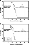
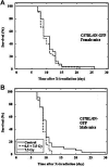
Application of green fluorescent protein (GFP) in a variety of biosystems as a unique bioindicator or biomarker has revolutionized biological research and made groundbreaking achievements, while increasing evidence has shown alterations in biological properties and physiological functions of the cells and animals overexpressing transgenic GFP. In this work, response to total body irradiation (TBI) was comparatively studied in GFP transgenic C57BL/6-Tg (CAG-EGFP) mice and C57BL/6 N wild type mice. It was demonstrated that GFP transgenic mice were more sensitive to radiation-induced bone marrow death, and no adaptive response could be induced. In the nucleated bone marrow cells of GFP transgenic mice exposed to a middle dose, there was a significant increase in both the percentage of cells expressing pro-apoptotic gene Bax and apoptotic cell death. While in wild type cells, lower expression of pro-apoptotic gene Bax and higher expression of anti-apoptotic gene Bcl-2, and significant lower induction of apoptosis were observed compared to GFP transgenic cells. Results suggest that presence of GFP could alter response to TBI at whole body, cellular and molecular levels in mice. These findings indicate that there could be a major influence on the interpretation of the results obtained in GFP transgenic mice.
Keywords: GFP transgenic mice; adaptive response; apoptosis; bone marrow death; green fluorescent protein; ionizing radiation.
© The Author(s) 2020.
Conflict of interest statement
Declaration of Conflicting Interests: The author(s) declared no potential conflicts of interest with respect to the research, authorship, and/or publication of this article.
Figures


Similar articles
-
Altered Induction of Reactive Oxygen Species by X-rays in Hematopoietic Cells of C57BL/6-Tg (CAG-EGFP) Mice.Int J Mol Sci. 2021 Jun 28;22(13):6929. doi: 10.3390/ijms22136929. Int J Mol Sci. 2021. PMID: 34203224 Free PMC article.
-
Novel enhanced GFP-positive congenic inbred strain establishment and application of tumor-bearing nude mouse model.Cancer Sci. 2020 Oct;111(10):3626-3638. doi: 10.1111/cas.14545. Epub 2020 Aug 11. Cancer Sci. 2020. PMID: 32589305 Free PMC article.
-
Bone mass and microarchitecture of irradiated and bone marrow-transplanted mice: influences of the donor strain.Osteoporos Int. 2009 Mar;20(3):435-43. doi: 10.1007/s00198-008-0658-3. Epub 2008 Jun 12. Osteoporos Int. 2009. PMID: 18548305
-
Contribution of different bone marrow-derived cell types in endometrial regeneration using an irradiated murine model.Fertil Steril. 2015 Jun;103(6):1596-605.e1. doi: 10.1016/j.fertnstert.2015.02.030. Epub 2015 Mar 23. Fertil Steril. 2015. PMID: 25813284
-
Suppression of apoptosis by bcl-2 overexpression in lymphoid cells of transgenic zebrafish.Blood. 2005 Apr 15;105(8):3278-85. doi: 10.1182/blood-2004-08-3073. Epub 2004 Dec 23. Blood. 2005. PMID: 15618471
Cited by
-
Recombinant cell-detecting RaDR-GFP in mice reveals an association between genomic instability and radiation-induced-thymic lymphoma.Am J Cancer Res. 2022 Feb 15;12(2):562-573. eCollection 2022. Am J Cancer Res. 2022. PMID: 35261787 Free PMC article.
-
Altered Induction of Reactive Oxygen Species by X-rays in Hematopoietic Cells of C57BL/6-Tg (CAG-EGFP) Mice.Int J Mol Sci. 2021 Jun 28;22(13):6929. doi: 10.3390/ijms22136929. Int J Mol Sci. 2021. PMID: 34203224 Free PMC article.
-
An exploratory approach to identify microRNAs as circulatory biomarker candidates for epilepsy-associated psychiatric comorbidities in an electrical post-status epilepticus model.Sci Rep. 2023 Mar 20;13(1):4552. doi: 10.1038/s41598-023-31017-9. Sci Rep. 2023. PMID: 36941269 Free PMC article.
References
-
- Shimomura O. Discovery of green fluorescent protein (GFP) (Nobel Lecture). Angew Chem Int Ed Engl. 2009;48(31):5590–5602. - PubMed
-
- Sarkar P, Chattopadhyay A. GFP fluorescence: a few lesser-known nuggets that make it work. J Biosci. 2018;43(3):421–430. - PubMed
-
- Tsien RY. The green fluorescent protein. Annu Rev Biochem. 1998;67:509–544. - PubMed
LinkOut - more resources
Full Text Sources
Research Materials
Miscellaneous

