Review
. 2020 Aug 14:13:156.
doi: 10.3389/fnmol.2020.00156. eCollection 2020.

Pathomechanisms and Clinical Implications of Myasthenic Syndromes Exacerbated and Induced by Medical Treatments

Affiliations
Review

Pathomechanisms and Clinical Implications of Myasthenic Syndromes Exacerbated and Induced by Medical Treatments

Martin Krenn et al. Front Mol Neurosci. .

Abstract

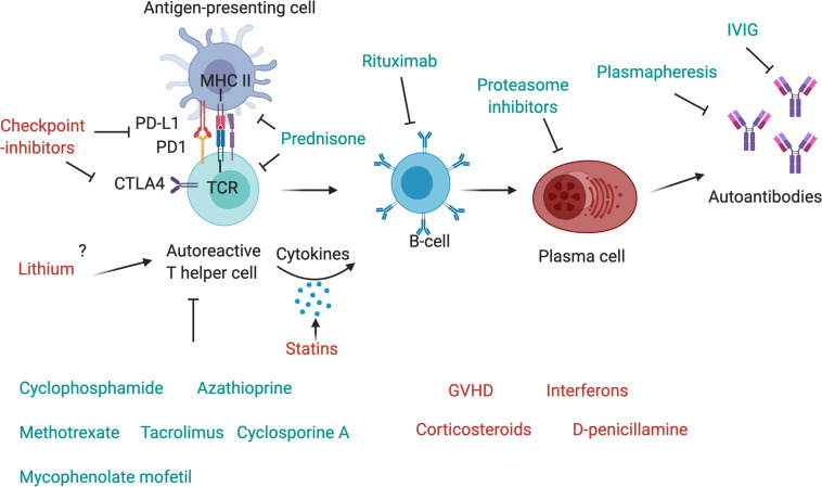
Myasthenic syndromes are typically characterized by muscle weakness and increased fatigability due to an impaired transmission at the neuromuscular junction (NMJ). Most cases are caused by acquired autoimmune conditions such as myasthenia gravis (MG), typically with antibodies against the acetylcholine receptor (AChR). Different drugs are among the major factors that may complicate pre-existing autoimmune myasthenic conditions by further impairing transmission at the NMJ. Some clinical observations are substantiated by experimental data, indicating that presynaptic, postsynaptic or more complex pathomechanisms at the NMJ may be involved, depending on the individual compound. Most robust data exist for the risks associated with some antibiotics (e.g., aminoglycosides, ketolides, fluoroquinolones) and cardiovascular medications (e.g., class Ia antiarrhythmics, beta blockers). Apart from primarily autoimmune-mediated disorders of the NMJ, de novo myasthenic manifestations may also be triggered by medical treatments that induce an autoimmune reaction. Most notably, there is growing evidence that the immune checkpoint inhibitors (ICI), a modern class of drugs to treat various malignancies, represent a relevant risk factor to develop severe and progressive medication-induced myasthenia via an immune-mediated mechanism. From a clinical perspective, it is of utmost importance for the treating physicians to be aware of such adverse treatment effects and their consequences. In this article, we aim to summarize existing evidence regarding the key molecular and immunological mechanisms as well as the clinical implications of medication-aggravated and medication-induced myasthenic syndromes.

Keywords: drug-induced myasthenia; drug-related myasthenia; immune checkpoint inhibitors; neuromuscular junction; neuromuscular transmission.


Figures

FIGURE 1
FIGURE 1
Drugs and antibodies that affect the function of the neuromuscular junction. Upon arrival of an action potential, presynaptic P-type VGCC (voltage-gated calcium channels) open and calcium enters the motoneuron terminal. This may be negatively affected by autoantibodies that block the VGCC, Mg+ or Verapamil. Calcium stimulates Ca2+ mediated exocytosis of neurotransmitters via the SNARE complex, which is negatively affected by BoNT-A which induces cleavage of one of its components, SNAP-25. Release of acetylcholine may also be reduced by corticosteroids or antibiotics. At the postsynaptic side, AChRs are densely aggregated, which is regulated via the agrin-Lrp4-MuSK-Dok-7 signaling pathway. Neural agrin is released by the motoneuron and binds to its co-receptor, Lrp4, located in the muscle membrane. Lrp4 and agrin bind as a dimer of dimers to the muscle-specific kinase (MuSK), a receptor tyrosine kinase that then phosphorylates itself, leading to the attachment of the adapter protein Dok-7. This binding leads to a full activation of MuSK and phosphorylation of downstream molecules, cumulating in the phosphorylation and dense aggregation of AChRs. The interaction between MuSK and Lrp4 can be blocked by autoantibodies, leading to interrupted signal transduction and reduced AChR clustering. The AChR clustering can be strengthened by SHP2-inhibitors and β2-adrenergic agonists, and de-stabilized by β2-adrenergic antagonists. Binding of acetylcholine to the AChR leads to opening of the channel, influx of cations, depolarization of the membrane and opening of voltage-gated Na + channels, which amplifies the depolarization. A range of drugs was shown to block the AChR, including fluoroquinolones, chloroquine, antibiotics, anesthetics, NMBA, telithromycin, curare and class Ia antiarrhythmics. Quinidine, a class Ia antiarrhythmic drug, may also be beneficial in the context of slow channel CMS with a pathological prolonged open state of the AChR. Voltage-gated Na+ channels are inhibited by phenytoin and class Ia antiarrhythmics. Acetylcholine is recycled in the synaptic cleft by the acetylcholinesterase (AChE), which hydrolyses acetylcholine to choline and acetate. AChE is arranged in tetramers attached to collagen q like tail (ColQ), which in turn is anchored to the synapse via binding to MuSK. AChE is inhibited by pyridostigmine, leading to prolonged presence of acetylcholine in the synaptic cleft, and is used as first line symptomatic treatment in myasthenia gravis.
FIGURE 2
FIGURE 2
Drugs affecting components of the immune system. The T-cell receptor (TCR) of autoreactive T-helper cells recognizes antigen peptides that are presented by the MHC II complex on antigen-presenting cells. The binding of programmed cell death protein 1 (PD-1) to its ligand, PD-L1, expressed, respectively, on antigen-presenting cell and T-cell, inhibits immune activation and is an important immune checkpoint to guard against autoimmunity. CTLA4 is another checkpoint that is expressed on regulatory T-cells and upregulated on conventional T cells after activation and turns off T-cell activation when it interacts with its ligands, CD80 or CD86, on antigen-presenting cells. Checkpoint inhibitors are used as therapy to stimulate an attack on tumor cells, but they may also activate autoreactive T-cells and induce autoimmunity. Corticosteroids such as Prednisone inhibit antigen processing and presenting in antigen-presenting cells and block T-cell activation in the nucleus. T-cells are further targeted by other immunosuppressant drugs, such as cyclophosphamide, azathioprine, methotrexate, mycophenolate mofetil, cyclosporin A or tacrolimus. T helper cells activate autoreactive B-cells via MHC II-antigen-TCR interaction and the secretion of pro-inflammatory cytokines, these may be increased by statins. B-cells can be depleted by the use of Rituximab, a therapeutic monoclonal antibody against CD20, which is beneficial in many antibody-mediated autoimmune diseases. Plasma cells, which produce autoantibodies, may be targeted by proteasome inhibitors, and autoantibodies can be depleted by plasmapheresis. IVIg are also a therapeutic drug in autoimmune diseases that ameliorate antibody-mediated effects, although their exact mechanism of action is unknown.

Similar articles

Cited by

References

    1. Abramsky O., Aharonov A., Teitelbaum D., Fuchs S. (1975). Myasthenia gravis and acetylcholine receptor. Effect of steroids in clinical course and cellular immune response to acetylcholine receptor. Arch. Neurol. 32 684–687. 10.1001/archneur.1975.00490520054008 - DOI - PubMed
    1. Adelman H. M., Winters P. R., Mahan C. S., Wallach P. M. (1995). D-penicillamine-induced myasthenia gravis: diagnosis obscured by coexisting chronic obstructive pulmonary disease. Am. J. Med. Sci. 309 191–193. 10.1097/00000441-199504000-00001 - DOI - PubMed
    1. Albers J. W., Beals C. A., Levine S. P. (1981). Neuromuscular transmission in rheumatoid arthritis, with and without penicillamine treatment. Neurology 31 1562–1564. 10.1212/wnl.31.12.1562 - DOI - PubMed
    1. Aldrich M. S., Kim Y. I., Sanders D. B. (1979). Effects of D-penicillamine on neuromuscular transmission in rats. Muscle Nerve 2 180–185. 10.1002/mus.880020305 - DOI - PubMed
    1. Alfrey A. C., Terman D. S., Brettschneider L., Simpson K. M., Ogden D. A. (1970). Hypermagnesemia after renal homotransplantation. Ann. Intern. Med. 73 367–371. 10.7326/0003-4819-73-3-367 - DOI - PubMed

LinkOut - more resources