

Knockdown of Death-Associated Protein Expression Induces Global Transcriptome Changes in Proliferating and Differentiating Muscle Satellite Cells
- PMID: 32922311
- PMCID: PMC7457014
- DOI: 10.3389/fphys.2020.01036
Knockdown of Death-Associated Protein Expression Induces Global Transcriptome Changes in Proliferating and Differentiating Muscle Satellite Cells
Abstract
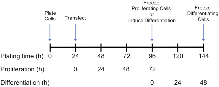
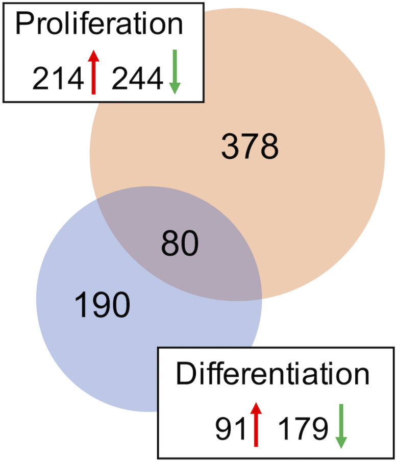
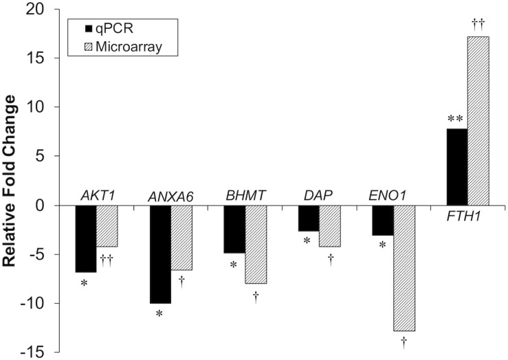
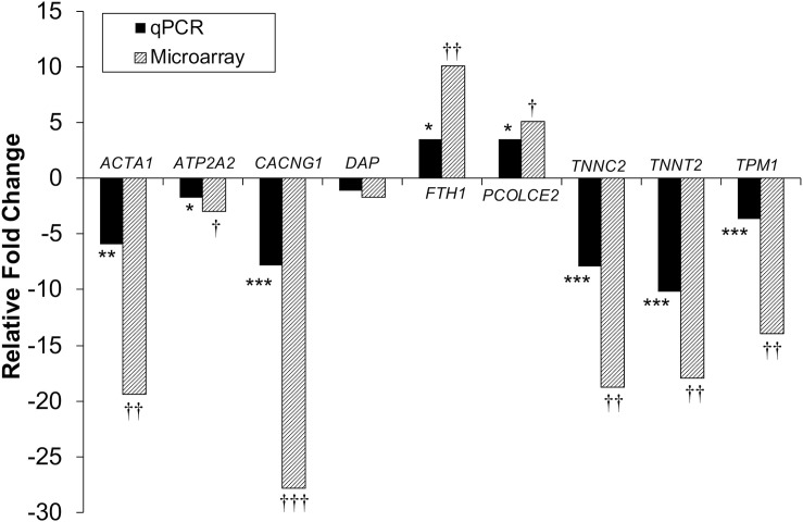
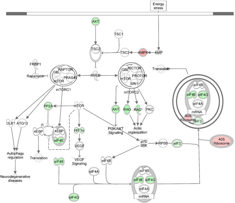
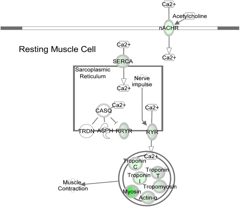
Death-associated protein (DAP) undergoes substantial changes in expression during turkey skeletal muscle development, decreasing from the 18 day embryonic stage to 1 day posthatch, and again from 1 day posthatch to 16 weeks of age. These changes suggest that DAP plays an important role at critical stages of the developmental process. The objective of this study was to elucidate the role of DAP in muscle development by examining the effect of reduced DAP expression on global gene expression in proliferating and differentiating turkey pectoralis major muscle satellite cells. Small interfering RNA was used to knock down expression of DAP and the transcriptome was subsequently profiled using a turkey skeletal muscle long oligonucleotide microarray. Microarray data were corroborated using quantitative real-time PCR. In proliferating cells, 458 loci, resulting in 378 uniquely annotated genes, showed differential expression (false discovery rate, FDR < 0.05). Pathway analysis highlighted altered eukaryotic translational initiation factors (eIFs) signaling, protein ubiquitination, sirtuin signaling, and mechanistic target of rapamycin (mTOR) signaling as the primary pathways affected in the knockdown proliferating cells. The findings underpinned the potential DAP involvement in cell proliferation of turkey satellite cells through the coordination between protein synthesis and cell cycle. In differentiating cells, 270 loci, accounting for 189 unique genes, showed differential expression (FDR < 0.05). Decreased expression of genes encoding various myofibrillar proteins and proteins involved in sarcoplasmic reticulum calcium flux suggests that DAP may affect regulation of calcium homeostasis and cytoskeleton signaling. This study provides the first evidence that reduced expression of DAP significantly alters the transcriptome profile of pectoralis major muscle satellite cells, thereby reducing proliferation and differentiation.
Keywords: death-associated protein; global gene expression; microarray; muscle; muscle development; satellite cells; turkey.
Copyright © 2020 Horton, Sporer, Tempelman, Malila, Reed, Velleman and Strasburg.
Figures
Similar articles
-
Versican, matrix Gla protein, and death-associated protein expression affect muscle satellite cell proliferation and differentiation.Poult Sci. 2012 Aug;91(8):1964-73. doi: 10.3382/ps.2012-02147. Poult Sci. 2012. PMID: 22802192
-
Response of Turkey Muscle Satellite Cells to Thermal Challenge. II. Transcriptome Effects in Differentiating Cells.Front Physiol. 2017 Nov 30;8:948. doi: 10.3389/fphys.2017.00948. eCollection 2017. Front Physiol. 2017. PMID: 29249977 Free PMC article.
-
Response of turkey muscle satellite cells to thermal challenge. I. transcriptome effects in proliferating cells.BMC Genomics. 2017 May 6;18(1):352. doi: 10.1186/s12864-017-3740-4. BMC Genomics. 2017. PMID: 28477619 Free PMC article.
-
Characterisation of equine satellite cell transcriptomic profile response to β-hydroxy-β-methylbutyrate (HMB).Br J Nutr. 2016 Oct;116(8):1315-1325. doi: 10.1017/S000711451600324X. Epub 2016 Oct 3. Br J Nutr. 2016. PMID: 27691998 Free PMC article.
-
Death associated proteins (DAPs): from gene identification to the analysis of their apoptotic and tumor suppressive functions.Oncogene. 1998 Dec 24;17(25):3331-40. doi: 10.1038/sj.onc.1202588. Oncogene. 1998. PMID: 9916995 Review.
Cited by
-
From discovery to application: merging modern omics with traditional hypothesis-driven approaches in muscle myopathy studies.Front Physiol. 2025 Jan 20;15:1520196. doi: 10.3389/fphys.2024.1520196. eCollection 2024. Front Physiol. 2025. PMID: 39902467 Free PMC article. No abstract available.
-
Optimization of the seat position for a personal vehicle equipped with a crankset: pilot study.Sci Rep. 2024 Mar 9;14(1):5822. doi: 10.1038/s41598-024-56446-y. Sci Rep. 2024. PMID: 38461198 Free PMC article.
References
LinkOut - more resources
Full Text Sources
Molecular Biology Databases
Miscellaneous