. 2020 Aug 14:11:1771.
doi: 10.3389/fimmu.2020.01771. eCollection 2020.

Isatuximab Acts Through Fc-Dependent, Independent, and Direct Pathways to Kill Multiple Myeloma Cells

Affiliations

Isatuximab Acts Through Fc-Dependent, Independent, and Direct Pathways to Kill Multiple Myeloma Cells

Chen Zhu et al. Front Immunol. .

Abstract

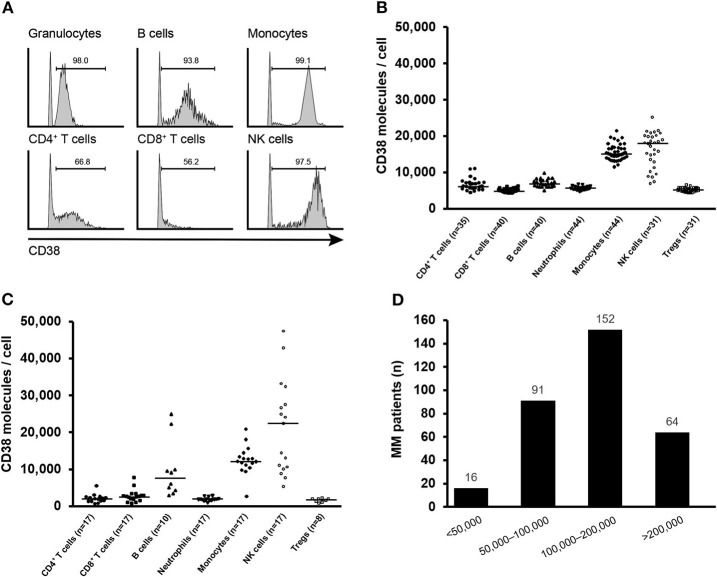
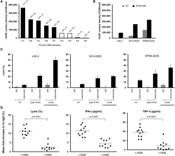
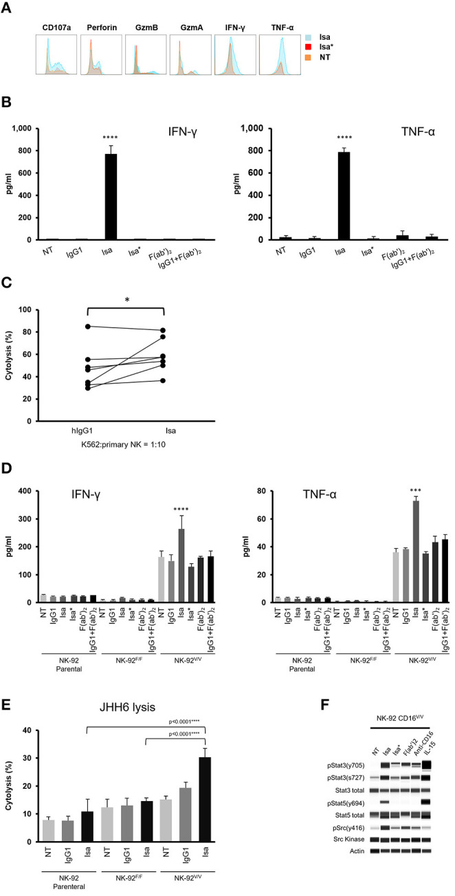
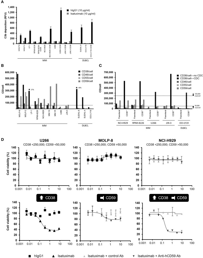
Isatuximab is a monoclonal antibody targeting the transmembrane receptor and ectoenzyme CD38, a protein highly expressed on hematological malignant cells, including those in multiple myeloma (MM). Upon binding to CD38-expressing MM cells, isatuximab is thought to induce tumor cell killing via fragment crystallizable (Fc)-dependent mechanisms, including antibody-dependent cellular cytotoxicity (ADCC), antibody-dependent cellular phagocytosis (ADCP), and complement-dependent cytotoxicity (CDC), as well as via direct Fc-independent mechanisms. Here, these mechanisms of action were investigated in MM and diffuse large B-cell lymphoma (DLBCL) cell lines, as well as in peripheral blood mononuclear cells derived from healthy donors, and in MM patient-derived samples. Our findings show that isatuximab-mediated cytotoxicity occurred primarily via ADCC and ADCP in MM cell lines and via ADCC and apoptosis in DLBCL cell lines expressing high levels of CD38. We identified the programmed cell death-1/programmed cell death-ligand 1 (PD-1/PD-L1) pathway and MM cell-secreted transforming growth factor-beta (TGF-β) as tumor cell-related features that could suppress CD38-mediated ADCC. Furthermore, we established that isatuximab can directly activate natural killer (NK) cells and promote NK cell-mediated cytotoxicity via crosslinking of CD38 and CD16. Finally, isatuximab-induced CDC was observed in cell lines with high CD38 receptor density (>250,000 molecules/cell) and limited expression of inhibitory complement regulatory proteins (CD46, CD55, and CD59; <50,000 molecules/cell). Taken together, our findings highlight mechanistic insights for isatuximab and provide support for a range of combination therapy approaches that could be tested for isatuximab in the future.

Keywords: CD38; PD-1; TGF-β; antibody-dependent cellular cytotoxicity; antibody-dependent cellular phagocytosis; isatuximab; multiple myeloma; natural killer cells.


Figures

Figure 1
Figure 1
CD38 expression on human immune cells. (A) Representative histograms of CD38 expression on indicated immune cell populations of human PBMCs. Gates for CD38+ population were set relative to an isotype control. (B) Median CD38 RD of CD38+ CD4+ T cells, CD8+ T cells, B cells, neutrophils, monocytes, NK cells, and Tregs obtained from healthy donor PBMCs and (C) bone marrow from patients with MM. (D) CD38 receptor density distribution on myeloma cells from MM patients (n = 323). MM, multiple myeloma; NK, natural killer; PBMC, peripheral blood mononuclear cell; RD, receptor density.
Figure 2
Figure 2
Isatuximab-mediated cytotoxic activities in MM and DLBCL cell lines. CD38 RD (y-axis) was measured in a panel of 22 MM and 14 DLBCL cell lines. Bars show CD38 RD. Data are representative of at least two independent experiments. (A) Cell lines marked with stars were tested for their sensitivity to isatuximab-induced ADCC lysis by NK-92.FcγRIIIAV/V effector cells. Numbers on top of the bars represent the percentage of cell lysis by isatuximab-mediated ADCC and the fold change of cell lysis between isatuximab- and IgG1 isotype-mediated ADCC. Black solid bars indicate >30% target cell lysis and >2-fold increase in lysis during ADCC induction with isatuximab. The dotted line is the estimated threshold of CD38 RD required to trigger isatuximab-mediated ADCC. (B) Tumor cells and effector THP-1 cells were co-cultured in the presence of 1 μg/ml of isatuximab or isotype control hIgG1. Phagocytosis by effector THP-1 cells was quantified by flow cytometry. Numbers represent the percentage of phagocytosis in the presence of isatuximab and the fold increase in phagocytic activity induced by isatuximab compared with hIgG1-treated samples. Stars indicate the cell lines tested in the ADCP assay. Black solid bars represent tumor cells responsive to isatuximab-mediated ADCP, which is defined by >20% phagocytosis and a >2-fold increase in phagocytic activity with isatuximab- compared with hIgG1-treated samples. The dotted line indicates the suggested CD38 RD threshold of 100,000 molecules/cell. (C) Cells were treated with 10 μg/ml of isatuximab or hIgG1 to induce apoptosis. Stars indicate the cell lines used to evaluate isatuximab pro-apoptotic activity measured by annexin V staining. Black solid bars represent tumor cells where isatuximab treatment led to >10% apoptosis and >2-fold increased apoptosis compared with IgG treatment. ADCC, antibody-dependent cellular cytotoxicity; ADCP, antibody-dependent cellular phagocytosis; DLBCL, diffuse large B-cell lymphoma; (h)IgG1, (human) immunoglobulin G1; MM, multiple myeloma; RD, receptor density.
Figure 3
Figure 3
Effect of CD38 RD on isatuximab-induced ADCC against primary MM cells. (A) Nine primary MM patient tumor cells were tested for their sensitivity to isatuximab-induced ADCC. The percentage of ADCC lysis was measured by calcein AM release from the tumor cells and the fold change relative to the IgG1 isotype control-treated samples expressed on top of the bars. Black solid bars indicate >30% lysis and a >2-fold increase in cell lysis after treatment with isatuximab vs. IgG1 isotype control. (B) MM cells were treated with 2 μM of ATRA for 48 h. Mean (SD) CD38 RD was quantified for cells with (black bars) or without (gray bars) ATRA treatment (n = 3). (C) Indicated MM cells were pretreated with or without 2 μM ATRA for 48 h and mixed with NK-92.CD16V/V effector cells in the presence of 1 μg/ml isatuximab or IgG1 isotype control. Mean (SD) NK cell-mediated cytolytic activities were measured by calcein AM release (n = 3). Cells overexpressing CD38 were used as positive controls of isatuximab-mediated ADCC. (D) 16 MM and 5 DLBCL cell lines were treated with isatuximab or IgG1 isotype control (1 μg/ml) for 5 h. Mean fold increase in the percentage of target cell lysis, IFN-γ production, and TNF-α production is shown for target cells grouped by CD38 expression level, either above or below the estimated threshold for ADCC (1 × 10⁵ molecules/cell; n = 1 for each cell line). Significance assessed by two-tailed t-test. ADCC, antibody-dependent cellular cytotoxicity; ATRA, all-trans retinoic acid; DLBCL, diffuse large B-cell lymphoma; IFN-γ, interferon-gamma; IgG1, immunoglobulin G1; MM, multiple myeloma; NK, natural killer; RD, receptor density; SD, standard deviation; TNF-α, tumor necrosis factor-alpha.
Figure 4
Figure 4
Effect of PD-L1/PD-1 interaction on isatuximab ADCC activity. (A) 5 × 10⁶ PBMCs and 5 × 10⁵ U266 cells overexpressing CD38 (U266.CD38++) were co-cultured in ultra-low attachment six-well plates for 7 days. The pre-conditioned PBMCs were then mixed with fresh U266.CD38++ cells with the presence of indicated concentrations of isatuximab or IgG1 isotype control at 37°C for 2–3 h to induce ADCC. Live CD138+ cells were quantified by flow cytometry. The percentages of live target cells were calculated relative to those for the IgG1 control. Each treatment was analyzed in triplicate. Results are Mean (SD). (B) PBMCs were pre-conditioned by MM cells as described in (A). The cells were analyzed for expression of PD-1 on CD56+ cells (NK cells) and PD-L1 on CD14+ cells (monocytes) by flow cytometry on day 0 and day 6 of the co-culture (n = 4–6). (C) 5 × 10⁶ PBMCs and 5 × 10⁵ U266 cells overexpressing CD38 (U266.CD38++) were co-cultured in ultra-low attachment six-well plates or a transwell system to prevent direct contact between MM cells and PBMCs for 6 days. The cells were analyzed for expression of PD-1 on CD56+ cells (NK cells) by flow cytometry on day 0 and day 6 of the co-culture. (D) PBMCs were cultured with or without U266.CD38++ cells for 6 days as described in (A). The cells were subsequently mixed with fresh U266.CD38++ target cells in the presence of 1 μg/ml of isatuximab or IgG1 isotype control at 37°C for 2–3 h. For the cells that received co-treatment, 5 μg/ml of anti-PD-1 or anti-PD-L1 antibody was added 30 min before adding 1 μg/ml of isatuximab to trigger ADCC. Following isatuximab treatment, live CD138+ cells (U266.CD38++ cells) were quantified by flow cytometry. ADCC, antibody-dependent cellular cytotoxicity; IFN-γ, interferon-gamma; IgG1, immunoglobulin G1; IL-6, interleukin-6; MM, multiple myeloma; NK, natural killer; NT, not treated; PBMC, peripheral blood mononuclear cell; PD-1, programmed cell death-1; PD-L1, programmed cell death-ligand 1; PD-L2, programmed cell death-ligand 2; SD, standard deviation; TGF-β, transforming growth factor-beta.
Figure 5
Figure 5
Effect of TGF-β on isatuximab-induced ADCC activity. (A) 1 × 10⁶ MM cells were cultured in 100 μl of complete medium. Culture medium supernatant was collected on days 1, 2, and 3; TGF-β1 secretion was determined by MSD Human TGF-β1 Kit. TGF-β mRNA expression data from CCLE. (B) TGF-β neutralization, using an anti-TGF-β1,2,3 antibody, in JJN-3 tumor cell supernatants restored isatuximab-induced ADCC. A transwell system was used to co-culture 2 × 10⁶ purified NK cells with 6 × 10⁶ JJN-3 cells in the presence of 50 μg/ml of anti-TGF-β antibody or isotype control at 37°C for 90 h. To quantify isatuximab-triggered ADCC, 4 × 10⁴ calcein AM-labeled RPMI-8226 cells were mixed with 1.2 × 10⁵ NK cells in the presence of isatuximab or Isa* at indicated concentrations and calcein AM release quantified. Analyses were performed in triplicate. Results are mean (SD). Ab, antibody; ADCC, antibody-dependent cellular cytotoxicity; CCLE, Cancer Cell Line Encyclopedia; Isa*, isatuximab mutant unable to bind to CD38; mRNA, messenger ribonucleic acid; MSD, Meso Scale Diagnostics; NK, natural killer; SD, standard deviation; TGF-β1, transforming growth factor-beta 1.
Figure 6
Figure 6
Isatuximab activates NK cells by crosslinking CD38 and CD16. (A) PBMCs were treated with or without 1 μg/ml of Isa* or Isa. After 7 days, NK cell activity was analyzed by intracellular staining for the production of perforin, granzyme-A, granzyme-B, IFN-γ, and TNF-α. (B) 1 × 10⁶ cells/ml of purified human NK cells (n = 3) were cultured overnight with or without the presence of IgG1, Isa, Isa*, F(ab')2, and IgG1+F(ab')2. Cell culture supernatants were collected and analyzed for the production of IFN-γ and TNF-α by MSD. ****p < 0.001 by unpaired t-test (n = 3). (C) K562 cells were labeled with calcein AM and co-cultured with purified human NK cells (n = 7) at the ratio 1:10 in the presence of 1 μg/ml of IgG1 or Isa for 1 h and cytotoxicity measured by calcein AM release. The experiment was performed in technical triplicate, with statistical analysis by two-tailed paired t-test. *p < 0.05. (D) 1 × 10⁶ cells/ml of human NK-92 parental, NK-92.CD16F/F or NK92.CD16V/V cells (n = 3) were cultured overnight with or without the presence of 1 μg/ml of IgG1, Isa, Isa*, F(ab')2, or IgG1+F(ab')2. Culture supernatants were collected and assayed for the production of IFN-γ and TNF-α by MSD. ***p < 0.001 by unpaired t-test (n = 3). (E) Cytotoxicity against CD38 JHH6 cells (n = 3) was measured by calcein AM release. The experiment was conducted in technical triplicate. ****p < 0.0001 by unpaired t-test (n = 3). (F) NK92.CD16V/V cells were serum-starved overnight and treated with or without Isa, Isa*, F(ab')2, anti-CD16, IL-15 for 15 min prior to cell lysis. Western blotting was used to determine activation of the Src kinase, STAT3 and STAT5. All results are mean (SD). F(ab')2, F(ab')2 portion of isatuximab; GzmA, granzyme-A; GzmB, granzyme-B; IFN-γ, interferon-gamma; IgG1, immunoglobulin G1; Isa, isatuximab; Isa*, isatuximab mutant unable to bind to CD38; MSD, Meso Scale Diagnostics; NK, natural killer; NT, not treated; PBMC, peripheral blood mononuclear cell; TNF-α, tumor necrosis factor-alpha.
Figure 7
Figure 7
Effect of isatuximab on phagocytic activity of human monocytes (A) Human monocytic THP-1 cells (1 × 10⁶ cells/ml) were seeded overnight along with FITC-labeled latex-IgG beads in the presence or absence of 1 μg/ml of IgG1, Isa, Isa*, F(ab')2, IgG1+F(ab')2, or 2 μM of PMA. Fluorescence microscopic analysis of phagocytosis was followed by incubation of THP-1 cells with latex beads and antibody treatment to determine phagocytic activity (n = 3). ****p < 0.0001 by unpaired t-test (n = 3). (B) The phagocytosis assay described in (A) was repeated using titrations of the indicated treatments (n = 3). (C) Monocytes isolated from three healthy human donors were seeded overnight along with FITC-labeled latex-IgG beads in the presence of 1 μg/ml of IgG1 or Isa. The experiment was conducted in technical triplicate. Error bars are SD. Statistical tests performed using one-way ANOVA. F(ab')2, F(ab')2 portion of isatuximab; FITC, fluorescein isothiocyanate; IgG1, immunoglobulin G1; Isa, isatuximab; Isa*, isatuximab mutant unable to bind to CD38; PMA, phorbol myristate acetate.
Figure 8
Figure 8
Expression of CD38 and complement regulatory proteins impact susceptibility to isatuximab-induced CDC. (A) C3b deposition was quantified by flow cytometry on the surface of MM and DLBCL cell lines, either with natural CD38 RD >250,000 molecules/cell, or for cell lines with parental cell line CD38 RD <250,000 molecules/cell with and without engineering to overexpress CD38 above this threshold. ⋆ indicates CDC sensitive cell lines. (B) Cell surface expression levels of CD38 and the complement regulatory proteins CD46, CD55, and CD59 were determined for MM and DLBCL cell lines by flow cytometry. (C) Complement regulatory protein expression was not affected by overexpression of CD38 in MM and DLBCL cell lines resistant to isatuximab-mediated CDC, for parental cell lines expressing high levels of complement regulatory proteins. (D) Isatuximab-triggered CDC lysis of resistant MM cell lines required increased CD38 expression (>250,000 molecules/cell) and functional inhibition of complement regulatory proteins such as CD59. Experiments were repeated at least twice, with each treatment analyzed in duplicate. Results are mean (SD). Ab, antibody; CDC, complement-dependent cytotoxicity; DLBCL, diffuse large B-cell lymphoma; hIgG1, human immunoglobulin G1; MM, multiple myeloma; RFU, relative fluorescence units; SD, standard deviation.

Similar articles

Cited by

References

    1. van de Donk NWCJ, Janmaat ML, Mutis T, Lammerts van Bueren JJ, Ahmadi T, Sasser AK, et al. Monoclonal antibodies targeting CD38 in hematological malignancies and beyond. Immunol Rev. (2016) 270:95–112. 10.1111/imr.12389 - DOI - PMC - PubMed
    1. Malavasi F, Deaglio S, Funaro A, Ferrero E, Horenstein AL, Ortolan E, et al. Evolution and function of the ADP ribosyl cyclase/CD38 gene family in physiology and pathology. Physiol Rev. (2008) 88:841–86. 10.1152/physrev.00035.2007 - DOI - PubMed
    1. Deaglio S, Morra M, Mallone R, Ausiello CM, Prager E, Garbarino G, et al. Human CD38 (ADP-ribosyl cyclase) is a counter-receptor of CD31, an Ig superfamily member. J Immunol. (1998) 160:395–402. - PubMed
    1. Muñoz P, Mittelbrunn M, de la Fuente H, Pérez-Martínez M, García-Pérez A, Ariza-Veguillas A, et al. Antigen-induced clustering of surface CD38 and recruitment of intracellular CD38 to the immunologic synapse. Blood. (2008) 111:3653–64. 10.1182/blood-2007-07-101600 - DOI - PubMed
    1. Krejcik J, Casneuf T, Nijhof IS, Verbist B, Bald J, Plesner T, et al. Daratumumab depletes CD38+ immune regulatory cells, promotes T-cell expansion, and skews T-cell repertoire in multiple myeloma. Blood. (2016) 128:384–94. 10.1182/blood-2015-12-687749 - DOI - PMC - PubMed

Publication types

MeSH terms

Substances