. 2020 Aug 14:11:2042.
doi: 10.3389/fimmu.2020.02042. eCollection 2020.

Methotrexate Enhances Apoptosis of Transmembrane TNF-Expressing Cells Treated With Anti-TNF Agents

Affiliations

Methotrexate Enhances Apoptosis of Transmembrane TNF-Expressing Cells Treated With Anti-TNF Agents

Qiaolei Wang et al. Front Immunol. .

Abstract

Background: Concomitant use of methotrexate (MTX) improves the clinical efficacy of anti-TNF agents in the treatment of rheumatoid arthritis (RA). We aimed to clarify the cytotoxic effect of MTX on transmembrane TNF (tmTNF)-expressing cells treated with anti-TNF agents.

Methods: Jurkat T cells stably expressing tmTNF were used for the following experiments. Cytotoxicity induced by an anti-TNF agent (infliximab, adalimumab, or certolizumab pegol) with concomitant MTX were compared with that by MTX alone or by an anti-TNF agent alone using flow cytometry. Apoptosis-induction mediated by reverse signal through tmTNF, complement-dependent cytotoxicity (CDC), antibody-dependent cell-mediated cytotoxicity (ADCC), and antibody-dependent cellular phagocytosis (ADCP) were evaluated. Folic acid and Y-27632, a Rho kinase inhibitor, were used as inhibitors to study intracellular signaling pathway in apoptosis induced by MTX and anti-TNF agents.

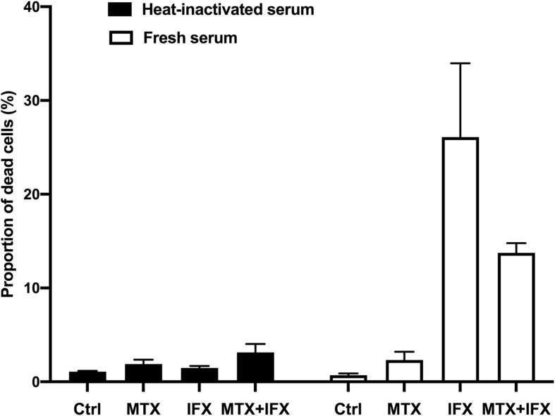
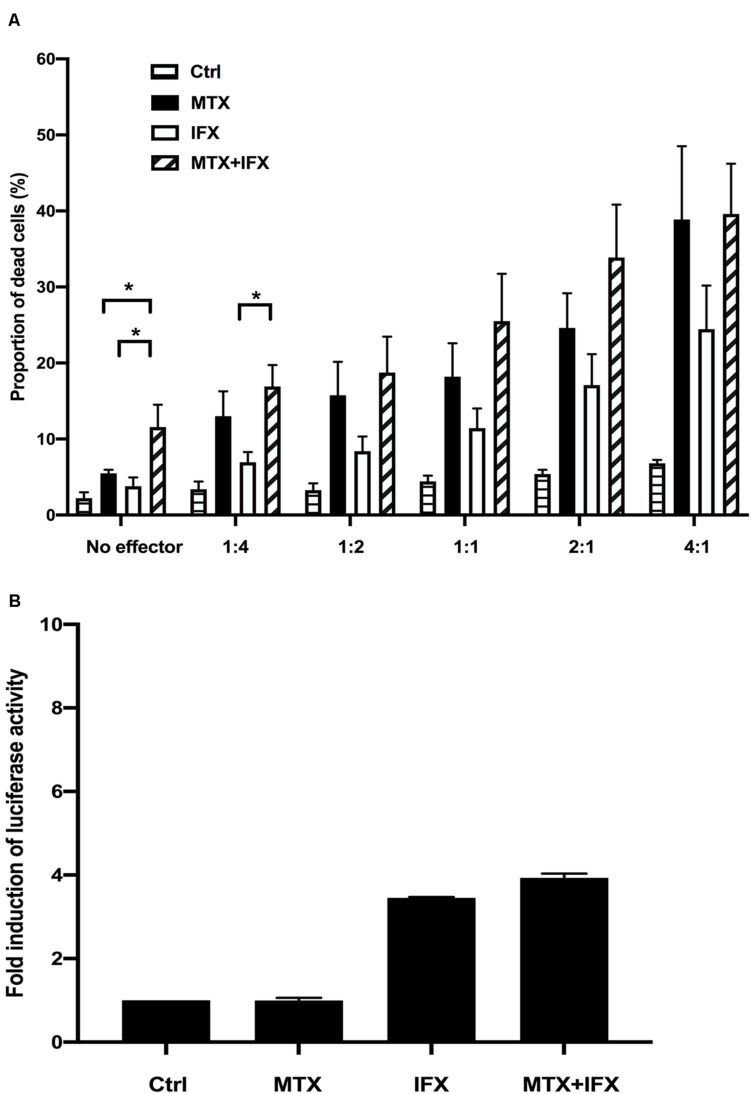
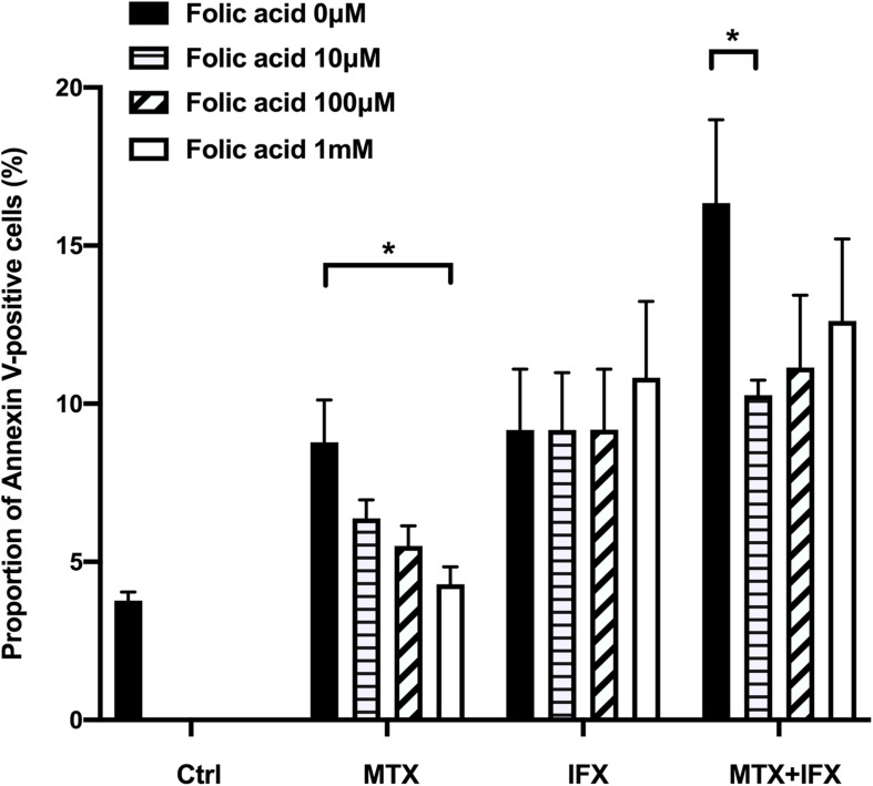
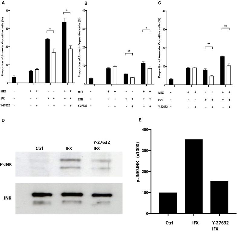
Results: Apoptosis of tmTNF-expressing cells was significantly increased by the concomitant administration of MTX and an anti-TNF agent, compared with MTX alone or an anti-TNF agent alone. The apoptosis induction by concomitant MTX was most pronounced in infliximab-treatment. Reverse signal transduction, but not CDC or ADCC/ADCP, was responsible for the coordinate effect of MTX and an anti-TNF agent on tmTNF-expressing cells. Folic acid inhibited MTX-mediated apoptosis, while Y-27632 suppressed JNK activation and infliximab-induced apoptosis via revere signal through tmTNF.

Conclusion: The apoptotic effect was enhanced by combination of MTX and an anti-TNF agent in tmTNF-expressing cells. The intracellular pathways induced by MTX and anti-TNF agents seem to be independent. These findings might explain at least in part improved the clinical response upon co-therapy of MTX and an anti-TNF agent in RA.

Keywords: anti-TNF agent; apoptosis; cytotoxicity; methotrexate; rheumatoid arthritis; transmembrane TNF.


Figures

FIGURE 1
FIGURE 1
Concomitant treatment of MTX with an anti-TNF agent enhanced apoptosis of tmTNF-expressing cells. (A) Mock or tmTNF-transfected Jurkat cells were stained with FITC-conjugated mouse anti-TNF-α mAb or control mouse IgG and analyzed by flow cytometry. The histograms show the cell surface expression levels of tmTNF. (B) TmTNF-Jurkat cells were incubated with 0.1 μM of MTX for 20 h, 0.01 μM of infliximab (IFX) for 6 h, or combination of MTX and IFX for 6 h after MTX for 14 h. Stimulated cells were stained with FITC-conjugated Annexin V and propidium iodide (PI), and the apoptotic cells were detected by flow cytometry. (C) The proportion of Annexin V-positive apoptotic cells was indicated. Values are mean (±SEM) of each group (n = 5/group). (D) TmTNF-expressing Jurkat cells were incubated with 0.1 μM MTX for 24 h, 0.01 μM ETN for 12 h, or combination of MTX and ETN for 12 h after MTX for 12 h. The proportion of Annexin V-positive cells was indicated. Values are mean (±SEM) of each group (n = 6/group). (E) TmTNF-expressing Jurkat cells were incubated with 0.1 μM MTX for 24 h, 0.01 μM CZP for 12 h, or combination of MTX and CZP for 12 h after MTX for 12 h. The proportion of Annexin V-positive cells was indicated. Values are mean (±SEM) of each group (n = 6/group). *p < 0.05, **p < 0.01, Mann-Whitney U tests.
FIGURE 2
FIGURE 2
Complement-dependent cytotoxicity induced by IFX in co-stimulation with MTX. TmTNF-expressing Jurkat cells were stimulated with 0.1 μM MTX for 25 h, 0.01 μM IFX for 1 h, or combination of MTX and IFX for 1 h after MTX for 1 h, with 5% human fresh serum (white bar) or heat-inactivated serum (black bar). Treated cells were stained with PI and dead cells were detected by flow cytometry. The proportion of dead cells was indicated. Values are mean (±SEM) of each group (n = 3/group).
FIGURE 3
FIGURE 3
Antibody-dependent cell-mediated cytotoxicity and antibody-dependent cellular phagocytosis induced by IFX in co-stimulation with MTX. (A) TmTNF-expressing Jurkat cells were used as target cells and labeled with membrane dye PKH26. Peripheral blood mononuclear cells (PBMCs) were used as effector cells and co-incubated with target cells on flat-bottom plates. The ratios of effector cells and target cells was from 1:4 to 4:1. Cells were incubated with MTX for 24 h, IFX for 2 h, or combination of MTX and IFX for 2 h after MTX for 24 h. As a control, target cells were incubated with effector cells without MTX and IFX. Treated cells were stained with TO-PRO-3 iodide. TO-PRO-3 iodide-positive dead cells in PKH26-positive target cells were detected by flow cytometry. Proportion of dead cells in target cells was indicated. Values are mean ± SEM; n = 4/group. (B) TmTNF Jurkat cells (2 × 10⁴ in 150 μl/well) were co-incubated with FcγRIIa-H effector cells (2 × 10⁴ in 150 μl/well) and stimulated with 0.1 μM MTX, 0.01 μM IFX, or MTX+IFX for 6 h. Treated cells were cultured with luciferase assay buffer for 20 min and the luminous cells were detected by a luminometer. Values are mean ± SEM; n = 3/group. *p < 0.05, Mann-Whitney U tests.
FIGURE 4
FIGURE 4
MTX-induced apoptosis was mediated by folate cycle inhibition. TmTNF-expressing Jurkat cells were incubated with 0.1 μM MTX for 24 h, 0.01 μM IFX for 4 h or combination of MTX and IFX for 4 h after MTX for 20 h, in the presence of 0, 10, 100, and 1000 μM folic acid. Proportion of Annexin V-positive cells were indicated. Values are mean (±SEM) of each group (n = 4/group). *p < 0.05, Mann Whitney U tests.
FIGURE 5
FIGURE 5
Infliximab-induced apoptosis was mediated by activation of Rho kinase. (A) TmTNF-expressing Jurkat cells were incubated with 0.1 μM MTX for 20 h, 0.01 μM IFX for 6 h, or combination of MTX and IFX for 6 h after MTX for 14 h, in the presence (white bar) or absence (black bar) of 0.01 μM of Rho kinase inhibitor (Y-27632). Apoptotic cells were analyzed by flow cytometry using Annexin V staining. The proportion of Annexin V-positive cells was indicated. (B) TmTNF- expressing Jurkat cells were stimulated with 0.1 μM MTX for 24 h, 0.01 μM ETN for 12 h, or combination of MTX and ETN for 12 h after MTX for 12 h with or without Y-27632. (C) TmTNF-expressing Jurkat cells were stimulated with 0.1 μM MTX for 24 h, 0.01 μM CZP for 12 h, or combination of MTX and CZP for 12 h after MTX for 12 h with or without Y-27632. Values are mean ± SEM; n = 4/group in (A) and n = 5/group in (B,C). *p < 0.05, **p < 0.01, Mann Whitney U tests. (D,E) TmTNF-expressing Jurkat cells were stimulated with 0.01 μM IFX for 2 h in the presence (Rho-i + IFX) or absence (IFX) of 0.01 μM of Y-27632. Stimulated cells or unstimulated cells (Ctrl) were lysed in lysis buffer and the levels of phosphorylated JNK (p-JNK) was analyzed in western blotting (D). The ratio of p-JNK: total JNK is indicated in (E).
FIGURE 6
FIGURE 6
Concomitant treatment of MTX with an anti-TNF agent enhanced apoptosis of activated human CD4+ T cells. (A) CD4+ T cells were cultivated in the 96-well plate and activated by anti-CD3/28 Ab for 1 h. And then, cells were stimulated with 5 μg/ml PHA for 48 h. Cells were stained with FITC-conjugated anti-TNF and expression levels of TNF on the cells surface was detected by flow cytometry. (B) CD4+ T cells were untreated (Ctrl) or treated with PHA and stimulated with 1 μM MTX, 1 μM IFX, or combination of MTX and IFX for 48 h. The stimulated cells were stained with FITC-conjugated Annexin V and PI, and the apoptotic cells were detected by flow cytometry. (C,D) Similar experiments were performed with ETN (C) or CZP (D) instead of IFX. Values are mean ± SEM; n = 5/group, *p < 0.05, **p < 0.01, Paired t-test.

Similar articles

Cited by

References

    1. Bradley JR. TNF-mediated inflammatory disease. J Pathol. (2008) 214:149–60. 10.1002/path.2287 - DOI - PubMed
    1. Feldmann M, Maini RN. Anti-TNF alpha therapy of rheumatoid arthritis: what have we learned? Annu Rev Immunol. (2001) 19:163–96. 10.1146/annurev.immunol.19.1.163 - DOI - PubMed
    1. Steeland S, Libert C, Vandenbroucke RE. A new venue of TNF targeting. Int J Mol Sci. (2018) 19:1442. 10.3390/ijms19051442 - DOI - PMC - PubMed
    1. Horiuchi T, Mitoma H, Harashima S, Tsukamoto H, Shimoda T. Transmembrane TNF-alpha: structure, function and interaction with anti-TNF agents. Rheumatology. (2010) 49:1215–28. 10.1093/rheumatology/keq031 - DOI - PMC - PubMed
    1. Mitoma H, Horiuchi T, Tsukamoto H. Binding activities of infliximab and etanercept to transmembrane tumor necrosis factor-alpha. Gastroenterology. (2004) 126:934–5; author reply 5–6. - PubMed

Publication types

MeSH terms