

Prevention of excessive scar formation using nanofibrous meshes made of biodegradable elastomer poly(3-hydroxybutyrate- co-3-hydroxyvalerate)
- PMID: 32922720
- PMCID: PMC7448259
- DOI: 10.1177/2041731420949332
Prevention of excessive scar formation using nanofibrous meshes made of biodegradable elastomer poly(3-hydroxybutyrate- co-3-hydroxyvalerate)
Abstract
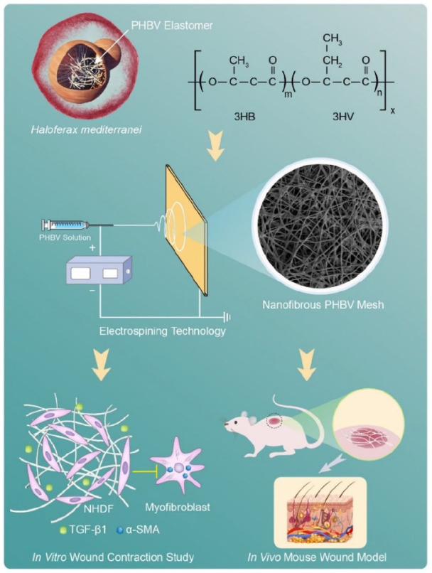
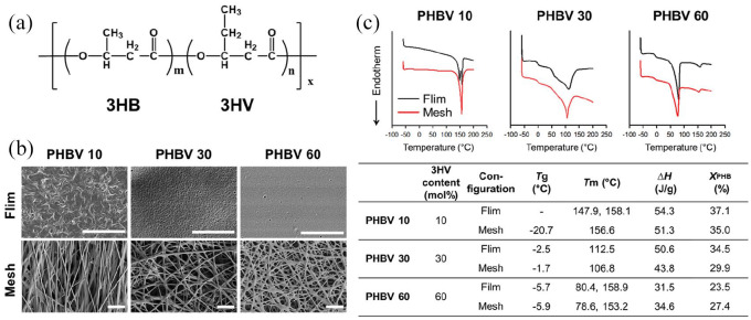
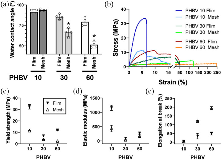
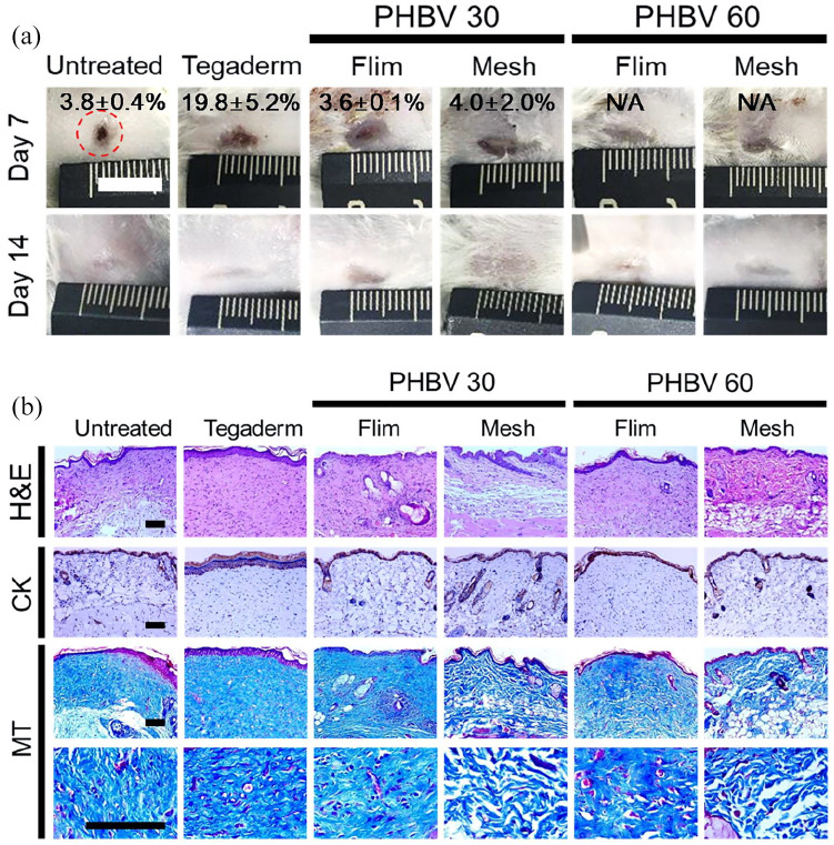
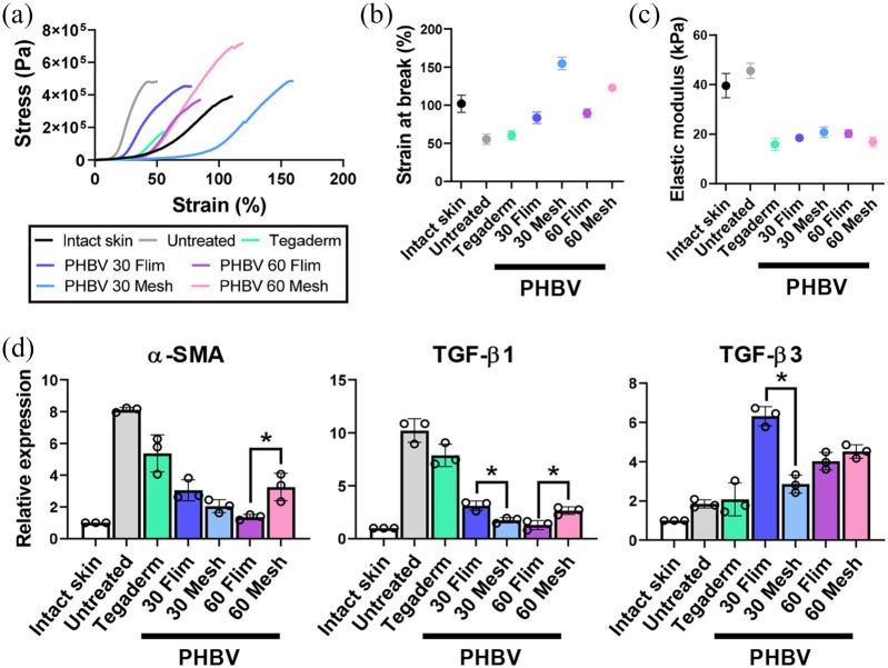
To reduce excessive scarring in wound healing, electrospun nanofibrous meshes, composed of haloarchaea-produced biodegradable elastomer poly(3-hydroxybutyrate-co-3-hydroxyvalerate) (PHBV), are fabricated for use as a wound dressing. Three PHBV polymers with different 3HV content are used to prepare either solution-cast films or electrospun nanofibrous meshes. As 3HV content increases, the crystallinity decreases and the scaffolds become more elastic. The nanofibrous meshes exhibit greater elasticity and elongation at break than films. When used to culture human dermal fibroblasts in vitro, PHBV meshes give better cell attachment and proliferation, less differentiation to myofibroblasts, and less substrate contraction. In a full-thickness mouse wound model, treatment with films or meshes enables regeneration of pale thin tissues without scabs, dehydration, or tubercular scar formation. The epidermis of wounds treated with meshes develop small invaginations in the dermis within 2 weeks, indicating hair follicle and sweat gland regeneration. Consistent with the in vitro results, meshes reduce myofibroblast differentiation in vivo through downregulation of α-SMA and TGF-β1, and upregulation of TGF-β3. The regenerated wounds treated with meshes are softer and more elastic than those treated with films. These results demonstrate that electrospun nanofibrous PHBV meshes mitigate excessive scar formation by regulating myofibroblast formation, showing their promise for use as wound dressings.
Keywords: Elastomer; PHBV; electrospun nanofiber; mechanical properties; scar formation.
© The Author(s) 2020.
Conflict of interest statement
Declaration of conflicting interests: The author(s) declared no potential conflicts of interest with respect to the research, authorship, and/or publication of this article.
Figures

References
-
- Aarabi S, Bhatt KA, Shi Y, et al. Mechanical load initiates hypertrophic scar formation through decreased cellular apoptosis. FASEB J 2007; 21: 3250–3261. - PubMed
-
- Lo DD, Zimmermann AS, Nauta A, et al. Scarless fetal skin wound healing update. Birth Defects Res C 2012; 96: 237–247. - PubMed
-
- Gurtner GC, Dauskardt RH, Wong VW, et al. Improving cutaneous scar formation by controlling the mechanical environment large animal and phase I studies. Ann Surg 2011; 254: 217–225. - PubMed
-
- Wang N, Tytell JD, Ingber DE. Mechanotransduction at a distance: mechanically coupling the extracellular matrix with the nucleus. Nat Rev Mol Cell Bio 2009; 10: 75–82. - PubMed
-
- Kadi A, Fawzi-Grancher S, Lakisic G, et al. Effect of cyclic stretching and TGF-beta on the SMAD pathway in fibroblasts. Bio-Med Mater Eng 2008; 18: S77–S86. - PubMed
LinkOut - more resources
Full Text Sources
Research Materials
Miscellaneous
