Case Reports
. 2020 Mar 20;6(3):20200015.
doi: 10.1259/bjrcr.20200015. eCollection 2020 Sep 1.

Non-traumatic complications of a solitary rib osteochondroma; an unusual cause of hemoptysis and pneumothorax

Affiliations
Case Reports

Non-traumatic complications of a solitary rib osteochondroma; an unusual cause of hemoptysis and pneumothorax

V E Versteegh et al. BJR Case Rep. .

Abstract

Osteochondromas are a very common and usually asymptomatic entity which may originate anywhere in the appendicular and axial skeleton. However, the ribs are a rare site of origin and here they may prove symptomatic for mechanical reasons. In this case report, we describe an unusual case of a symptomatic osteochondroma of the rib secondary to its location and unique shape, ultimately requiring surgical intervention.


Figures

Figure 1.
Figure 1.
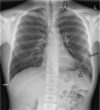
X-Thorax showing a leftsided apical pneumothorax (large arrow), left upper lobe consolidation (small arrow) and basal pleural adhesion (arrow head).
Figure 2.
Figure 2.
Axial CT angiography image (5 mm reconstruction; bone setting) showing osseous continuity of the osteochondroma and the sixth rib on the left side.
Figure 3.
Figure 3.
Sagittal and axial reconstruction CT angiography (in bone and pulmonary setting respectively), showing an osseous proliferation perforating the lung parenchyma causing a pneumothorax and pulmonary hemorrhage.
Figure 4.
Figure 4.
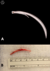
(a) 3D volume rendering reconstruction of CT angiography (Syngo.via, Siemens (Erlangen; Germany)) showing the osteochondroma in our patient originating from the 6th rib and its continuity with the underlying bone. (b) Post-operative specimen after resection of the osteochondroma. 3D, three-dimensional.
Figure 5.
Figure 5.
Per-operative view of our patient illustrating the osteochondroma compromising the thoracic cavity.
Figure 6.
Figure 6.
Per-operative view of our patient illustrating the pulmonary hemorrhage adjacent to the osteochondroma.

References

    1. Kitsoulis P, Galani V, Stefanaki K, Paraskevas G, Karatzias G, Agnantis NJ, et al. . Osteochondromas: review of the clinical, radiological and pathological features. In Vivo 2008; 22: 633–46. - PubMed
    1. Nam SJ, Kim S, Lim BJ, Yoon C-S, Kim TH, Suh J-S, Yoon C-S, Suh J-S, et al. . Imaging of primary chest wall tumors with radiologic-pathologic correlation. Radiographics 2011; 31: 749–70. doi: 10.1148/rg.313105509 - DOI - PubMed
    1. Waller DA, Newman RJ. Primary bone tumours of the thoracic skeleton: an audit of the Leeds regional bone tumour Registry. Thorax 1990; 45: 850–5. doi: 10.1136/thx.45.11.850 - DOI - PMC - PubMed
    1. Murphey MD, Choi JJ, Kransdorf MJ, Flemming DJ, Gannon FH. Imaging of osteochondroma: variants and complications with radiologic-pathologic correlation. Radiographics 2000; 20: 1407–34. doi: 10.1148/radiographics.20.5.g00se171407 - DOI - PubMed
    1. Limeme M, Mazhoud I, Zaghouani H, Laaribi M, Majdoub S, Amara H, et al. . Imaging of primary chest wall tumors. EPOS ECR, 2015 March 04-08; Vienna. Austria European Society of Radiology 2015;.

Publication types