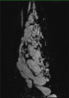
Novel use of arterial spin labelling in the imaging of peripheral vascular malformations
- PMID: 32922846
- PMCID: PMC7465750
- DOI: 10.1259/bjrcr.20200021
Novel use of arterial spin labelling in the imaging of peripheral vascular malformations
Abstract
We present a novel use of arterial spin labelling (ASL), a MRI perfusion technique, to assess a high-flow, peripheral vascular malformation (PVM), specifically a large arteriovenous malformation in the left forearm of a 20-year-old female. While there has been experience with ASL in the assessment of intracranial vascular malformations, there has been no known use of ASL in the evaluation of PVMs. We also discuss the potential benefits and limitations of ASL in the imaging of PVMs. The promising results from this case warrant further research on ASL in the investigation of PVMs.
© 2020 The Authors. Published by the British Institute of Radiology.
Figures


Similar articles
-
Feasibility of arterial spin labeling in evaluating high- and low-flow peripheral vascular malformations: a case series.BJR Case Rep. 2021 Aug 12;8(1):20210083. doi: 10.1259/bjrcr.20210083. eCollection 2022 Jan 1. BJR Case Rep. 2021. PMID: 35136637 Free PMC article.
-
Arterial spin labeling magnetic resonance imaging: toward noninvasive diagnosis and follow-up of pediatric brain arteriovenous malformations.J Neurosurg Pediatr. 2015 Apr;15(4):451-8. doi: 10.3171/2014.9.PEDS14194. Epub 2015 Jan 30. J Neurosurg Pediatr. 2015. PMID: 25634818
-
Association of developmental venous anomalies with perfusion abnormalities on arterial spin labeling and bolus perfusion-weighted imaging.J Neuroimaging. 2015 Mar-Apr;25(2):243-250. doi: 10.1111/jon.12119. Epub 2014 Apr 9. J Neuroimaging. 2015. PMID: 24717021
-
Mapping of cerebral perfusion territories using territorial arterial spin labeling: techniques and clinical application.NMR Biomed. 2013 Aug;26(8):901-12. doi: 10.1002/nbm.2836. Epub 2012 Jul 15. NMR Biomed. 2013. PMID: 22807022 Review.
-
Advances in arterial spin labelling MRI methods for measuring perfusion and collateral flow.J Cereb Blood Flow Metab. 2018 Sep;38(9):1461-1480. doi: 10.1177/0271678X17713434. Epub 2017 Jun 9. J Cereb Blood Flow Metab. 2018. PMID: 28598243 Free PMC article. Review.
Cited by
-
Feasibility of arterial spin labeling in evaluating high- and low-flow peripheral vascular malformations: a case series.BJR Case Rep. 2021 Aug 12;8(1):20210083. doi: 10.1259/bjrcr.20210083. eCollection 2022 Jan 1. BJR Case Rep. 2021. PMID: 35136637 Free PMC article.
References
Publication types
LinkOut - more resources
Full Text Sources

