

Case Reports
doi: 10.1259/bjrcr.20200075.
eCollection 2020 Sep 1.
A case of adrenal infarction in a patient with COVID 19 infection
Affiliations
- PMID: 32922854
- PMCID: PMC7465735
- DOI: 10.1259/bjrcr.20200075
Item in Clipboard
Case Reports
A case of adrenal infarction in a patient with COVID 19 infection
BJR Case Rep.
.
Abstract
This case report highlights an unusual presentation of acute adrenal infarction in a Covid-19 patient who presented with abdominal symptoms and hyponatraemia. We discuss the recent literature reviewing how Covid-19 creates a hypercoaguable state, with acute adrenal infarction as a possible prothrombotic complication.
© 2020 The Authors. Published by the British Institute of Radiology.
Figures
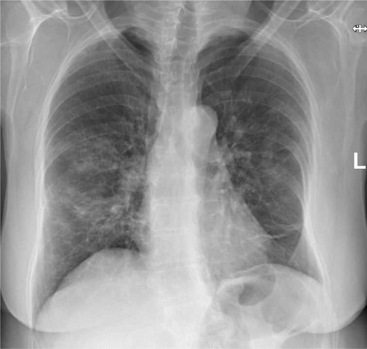
PA chest radiograph demonstrating bilateral, mid and lower zone, patchy parenchymal infiltrates in keeping with moderate Covid-19 pulmonary infection. PA, posteroanterior.
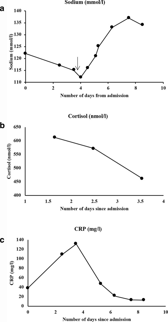
Trends of important blood parameters (sodium, cortisol and CRP) over the duration of the patient’s stay in hospital. The day of treatment commencement for the patient’s hyponatraemia has been demonstrated on the graph 2A (black arrow) with a clear subsequent improvement in the patient’s sodium levels. (Normal ranges: Sodium 133–146 mmol l−1, random cortisol expected to be >300 nmol l−1 and CRP 1–5 mg l−1).
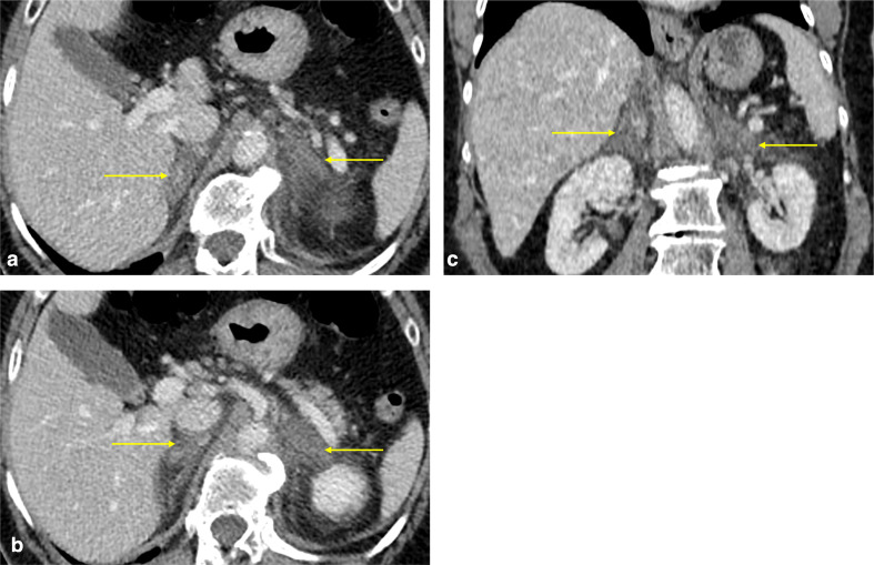
(a, b) Post-contrast axial and (c) coronal CT images of the abdomen on the portal venous phase demonstrating diffusely hypoattenuating, thickened adrenal glands with ill-defined contours and surrounding fat stranding (yellow arrows). Findings are in keeping with acute adrenal infarction.
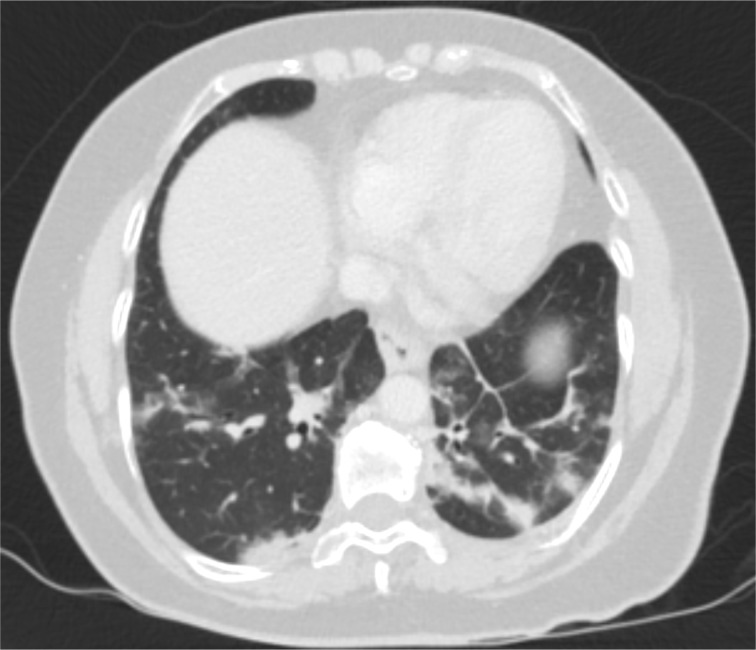
Post-contrast axial images of the CT abdomen and pelvis study at the level of the lung bases (lung window) demonstrating bilateral, peripheral areas of consolidation and ground glass opacities, as well as parenchymal bands. Findings are consistent with Covid-19 pulmonary infection.
References
Publication types
LinkOut - more resources
Full Text Sources
