Case Reports
. 2020 Jul 24;6(3):20200098.
doi: 10.1259/bjrcr.20200098. eCollection 2020 Sep 1.

Acute Flaccid Myelitis in COVID-19

Affiliations
Case Reports

Acute Flaccid Myelitis in COVID-19

Mohamed Abdelhady et al. BJR Case Rep. .

Abstract

Spinal cord imaging findings in COVID-19 are evolving with the increasing frequency of neurological symptoms among COVID-19 patients. Several mechanisms are postulated to be the cause of central nervous system affection including direct virus neuroinvasive potential, post infectious secondary immunogenic hyperreaction, hypercoagulability, sepsis and possible vasculitis as well as systemic and metabolic complications associated with critical illness. Only a few case reports of spinal cord imaging findings are described in COVID-19, which include transverse myelitis, acute disseminated encephalomyelitis and post-infectious Guillain Barre' syndrome. We are describing a case of myelitis which, to the best of our knowledge, is the first reported case of myelitis in COVID-19.


Figures

Figure 1.
Figure 1.
Frontal chest radiograph displays bilateral scattered lung infiltrates most pronounced in right lower lung zone.
Figure 2.
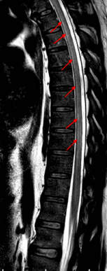
Figure 2.
Sagittal T2 weighted image MRI shows slight hyperintensities in the ventral aspect of the upper and mid-thoracic cord (arrows).
Figure 3.
Figure 3.
Axial T2 weighted images MRI at the level of upper thoracic cord (a, b) and at the level of mid thoracic cord (c, d) show hyperintensities in the ventral horns (arrows) of upper and mid thoracic cord bilaterally.
Figure 4.
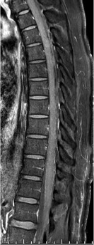
Figure 4.
Sagittal post-contrast fat-saturated T1 weighted image MRI shows no evidence of abnormal enhancement in the affected segments of the cord.

References

    1. Kincaid O, Lipton HL. Viral myelitis: an update. Curr Neurol Neurosci Rep 2006; 63: 469–74. doi: 10.1007/s11910-006-0048-1 - DOI - PubMed
    1. Talbott JF, Narvid J, Chazen JL, Chin CT, Shah V. An imaging-based approach to spinal cord infection. Seminars in Ultrasound, CT and MRI 2016; 37: 411–30. doi: 10.1053/j.sult.2016.05.006 - DOI - PubMed
    1. Petropoulou KA, Gordon SM, Prayson RA, Ruggierri PM. West Nile virus meningoencephalitis: MR imaging findings. American journal of neuroradiology 2005; 26: 1986–95PMID. - PMC - PubMed
    1. Lycklama G, Thompson A, Filippi M, Miller D, Polman C, Fazekas F, et al. . Spinal-Cord MRI in multiple sclerosis. The Lancet Neurology 2003; 2: 555–62. doi: 10.1016/S1474-4422(03)00504-0 - DOI - PubMed
    1. Kim HJ, Paul F, Lana-Peixoto MA, Tenembaum S, Asgari N, Palace J, et al. . Mri characteristics of neuromyelitis optica spectrum disorder: an international update. Neurology 2015; 84: 1165–73. - PMC - PubMed

Publication types