

Natural course of pineal cysts-a radiographic study
- PMID: 32922893
- PMCID: PMC7398253
- DOI: 10.1186/s41016-018-0142-7
Natural course of pineal cysts-a radiographic study
Abstract
Background: Pineal cysts (PCs) are a benign lesion of the pineal gland that have been known to the medical community for a long time. With a prevalence rate of approximately 1% in the general population, PC is often a reason for medical counseling. The natural course of PC morphology has not been well described. In this study, we present a longitudinal magnetic resonance imaging (MRI) study of patients with PCs, with special focus on those who showed an increase or decrease in PC size.
Methods: We enrolled all patients with a PC who were referred to our department between January 2000 and January 2018. Each patient underwent a clinical examination, and the patient's age, sex, and presenting signs and symptoms were noted. MRI was performed during periodic examinations, and a clinical and radiological course was reassessed.
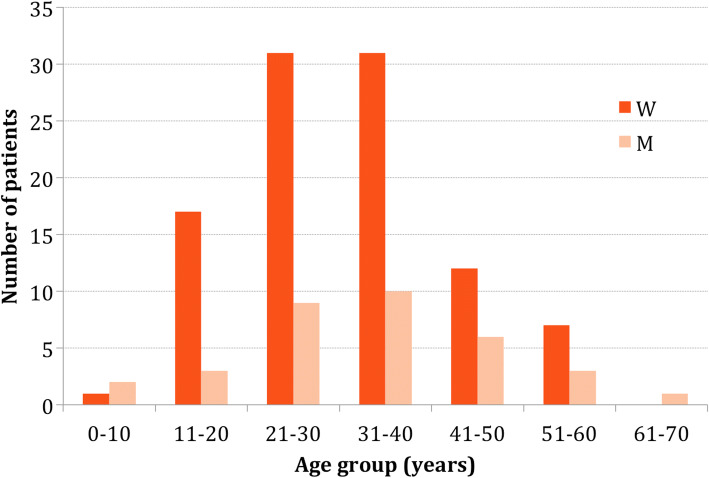
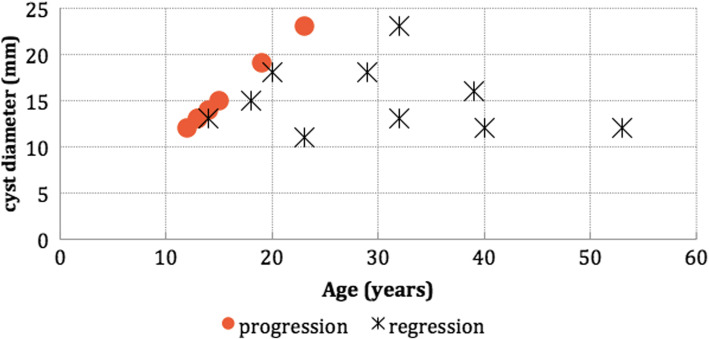
Results: In total, 133 patients (99 women, 34 men) were enrolled. The mean maximum diameter was 12.7 ± 5.2 mm (range 7-35 mm). PCs increased in size during the follow-up in seven patients (5.3%) and decreased in size in 10 (7.5%). The remaining cysts (n = 116, 87.2%) were stable over the follow-up period. Analyzing patients according to cyst size change, we found a significant difference in the mean age between the PC progression group and PC regression group (p = 0.01). The mean size of the PCs at the time of diagnosis did not differ significantly between the two groups (p = 0.81). We diagnosed two cases of pineal apoplexy.
Conclusion: We found that PCs are a dynamic structure that may change in size during the patient's lifetime. Patients with an increase in PC size were significantly younger than patients with a decrease in size. Therefore, PC growth in the first, second, and third decennium is normal and does not justify medical intervention. Surgery is indicated in cases of hydrocephalus and Parinaud's syndrome or in atypical cysts when neoplasia is suspected. The size of a PC does not predict PC behavior in terms of a future increase or decrease in size.
Keywords: Magnetic resonance imaging; Natural history; Neurosurgery; Pineal cyst.
© The Author(s) 2018.
Conflict of interest statement
Competing interestsThe authors declare that they have no competing interests.
Figures

Similar articles
-
Pineal cyst resection in the absence of ventriculomegaly or Parinaud's syndrome: clinical outcomes and implications for patient selection.J Neurosurg. 2015 Aug;123(2):352-6. doi: 10.3171/2014.9.JNS141081. Epub 2015 May 1. J Neurosurg. 2015. PMID: 25932610
-
Pineal cysts: Does anyone need long-term follow up?J Clin Neurosci. 2021 Jan;83:146-151. doi: 10.1016/j.jocn.2020.10.051. Epub 2020 Nov 30. J Clin Neurosci. 2021. PMID: 33272885
-
Evaluation of pineal cysts with magnetic resonance imaging.World J Radiol. 2018 Jul 28;10(7):65-77. doi: 10.4329/wjr.v10.i7.65. World J Radiol. 2018. PMID: 30079153 Free PMC article.
-
Pineal Apoplexy: A Case Series and Review of the Literature.J Neurol Surg A Cent Eur Neurosurg. 2022 Jan;83(1):31-38. doi: 10.1055/s-0041-1723813. Epub 2021 Jun 2. J Neurol Surg A Cent Eur Neurosurg. 2022. PMID: 34077982 Review.
-
[Pineal cyst].Zh Vopr Neirokhir Im N N Burdenko. 2017;81(4):113-120. doi: 10.17116/neiro2017814113-120. Zh Vopr Neirokhir Im N N Burdenko. 2017. PMID: 28914878 Review. Russian.
Cited by
-
Surgical strategy for symptomatic pineal cyst: is endoscopit third ventriculostomy necessary in addition to cyst fenestration?Nagoya J Med Sci. 2021 Aug;83(3):627-633. doi: 10.18999/nagjms.83.3.627. Nagoya J Med Sci. 2021. PMID: 34552294 Free PMC article.
-
Pineal cyst apoplexy and memory loss: a novel complication.Radiol Case Rep. 2022 Aug 5;17(10):3739-3744. doi: 10.1016/j.radcr.2022.07.055. eCollection 2022 Oct. Radiol Case Rep. 2022. PMID: 35965931 Free PMC article.
References
-
- Virchow R. Die Krankhaften Geschwülste. Berlin: Verlag von August Hirschwald; 1865.
-
- Campbell AW. Notes of two cases of dilatation of central cavity or ventricle of the pineal gland. Tr Path Soc. 1899;50:15–17.
-
- Pussep L. Die operative Entfernung einer Zyste der Glandula pinealis. Neurol Zentralbl. 1914;33:560–563.
-
- Bregant T, Rados M, Derganc M, Neubauer D, Kostovic I. Pineal cysts - a benign consequence of mild hypoxia in a near-term brain? Neuro Endocrinol Lett. 2011;32:663–666. - PubMed
LinkOut - more resources
Full Text Sources