. 2020 Jun 11;9(1):1777046.
doi: 10.1080/2162402X.2020.1777046.

Adoptive cell therapy of triple negative breast cancer with redirected cytokine-induced killer cells

Affiliations

Adoptive cell therapy of triple negative breast cancer with redirected cytokine-induced killer cells

Roberta Sommaggio et al. Oncoimmunology. .

Abstract

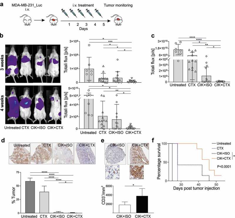
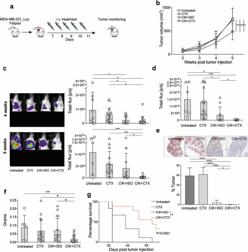
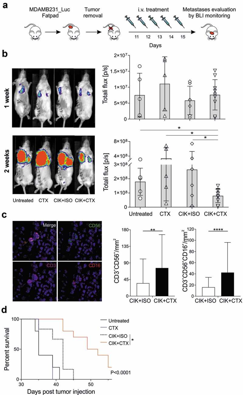
Cytokine-Induced Killer (CIK) cells share several functional and phenotypical properties of both T and natural killer (NK) cells. They represent an attractive approach for cell-based immunotherapy, as they do not require antigen-specific priming for tumor cell recognition, and can be rapidly expanded in vitro. Their relevant expression of FcγRIIIa (CD16a) can be exploited in combination with clinical-grade monoclonal antibodies (mAbs) to redirect their lytic activity in an antigen-specific manner. Here, we report the efficacy of this combined approach against triple negative breast cancer (TNBC), an aggressive tumor that still requires therapeutic options. Different primitive and metastatic TNBC cancer mouse models were established in NSG mice, either by implanting patient-derived TNBC samples or injecting MDA-MB-231 cells orthotopically or intravenously. The combined treatment consisted in the repeated intratumoral or intravenous injection of CIK cells and cetuximab. Tumor growth and metastasis were monitored by bioluminescence or immunohistochemistry, and survival was recorded. CIK cells plus cetuximab significantly restrained primitive tumor growth in mice, either in patient-derived tumor xenografts or MDA-MB-231 cell line models. Moreover, this approach almost completely abolished metastasis spreading and dramatically improved survival. The antigen-specific mAb favored tumor and metastasis tissue infiltration by CIK cells, and led to an enrichment of the CD16a+ subset.Data highlight the potentiality of this novel immunotherapy strategy where a nonspecific cytotoxic cell population can be converted into tumor-specific effectors with clinical-grade antibodies, thus providing not only a therapeutic option for TNBC but also a valid alternative to more complex approaches based on chimeric antigen receptor-engineered cells.

List of abbreviations: ACT, Adoptive Cell Transfer; ADCC, Antibody-Dependent Cell-mediated Cytotoxicity; ADP, Adenosine diphosphate; BLI, Bioluminescence Imaging; CAR, Chimeric Antigen Receptor; CIK, Cytokine Induced Killer cells; CTX, Cetuximab; DMEM, Dulbecco's Modified Eagle Medium; EGFR, Human Epidermal Growth Factor 1; ER, Estrogen; FBS, Fetal Bovine Serum; FFPE, Formalin-Fixed Paraffin-Embedded; GMP, Good Manufacturing Practices; GVHD, Graft Versus Host Disease; HER2, Human Epidermal Growth Factor 2; HRP, Horseradish Peroxidase; IFN-γ, Interferon-γ; IHC, Immunohistochemistry; IL-2, Interleukin-2; ISO, Irrelevant antibody; i.t., intratumoral; i.v., intravenous, mAbs, Monoclonal Antibodies; mIHC, Multiplex Fluorescence Immunohistochemistry; MHC, Major Histocompatibility Complex; NK, Natural Killer; NKG2D, Natural-Killer group 2 member D; NSG, NOD/SCID common γ chain knockout; PARP, Poly ADP-ribose polymerase; PBMCs, Peripheral Blood Mononuclear Cells; PBS, Phosphate-buffered saline; PDX, Patient-derived xenograft; PR, Progesterone; rhIFN-γ, Recombinant Human Interferon-γ; RPMI, Roswell Park Memorial Institute; STR, Short tandem Repeat; TCR, T Cell Receptor; TNBC, Triple Negative Breast Cancer; TSA, Tyramide Signal Amplification.

Keywords: Cytokine-induced killer cells (CIK); TNBC mouse models; adoptive cells therapy (ACT); cetuximab; triple negative breast cancer (TNBC).


Figures

Figure 1.
Figure 1.
CIK cell cytotoxicity against TNBC cell lines is enhanced when combined with CTX. (a) CIK cell bulk cultures were characterized by flow cytometry for the content of CD3+CD56+ and CD3+CD56+CD16a+ subsets, and CD3CD56+ cells. Dot plot of one representative donor is showed (upper panels). Histograms refer to the mean expression ± SD of 15 independent expansions from distinct healthy donors (lower panels). (b) MDA-MB-231 (left panel) and MDA-MB-468 (right panel) were analyzed for EGFR expression (pink) by flow cytometry. A fluorochrome-conjugated secondary antibody alone (gray) served as negative control. (c) CIK cells were assessed for cytotoxicity against MDA-MB-231 (left panel) and MDA-MB-468 (right panel) tumor cell lines. Lytic activity was measured in the absence (white boxes) or in the presence of an isotype control mAb (ISO, light gray boxes) or cetuximab (CTX, dark gray boxes) at an E/T ratio of 50:1; boxes indicate mean value ± SD of 15 independent experiments. ***P < .001, ****P < .0001.
Figure 2.
Figure 2.
CIK cells and Cetuximab combined treatment is therapeutically efficient against TNBC patient-derived xenografts. (a) Representative image of a TNBC patient-derived xenograft IHC-stained for EGFR (brown). (b) The experimental protocol consisted in the implantation of PDX in the fat pad area of NSG mice, followed by i.t. or i.v. daily treatments for five days, starting when tumors reached about 500 mm3. (c) Mice received i.t. CIK cells in combination with CTX (n = 16; inverted triangle) or ISO (n = 13; diamond), CTX alone (n = 10; triangle), or were left untreated (n = 7; circle). Tumor volume was measured by caliper measurement. Data are expressed as mean ± SD (Repeated measures ANOVA). (d) Representative images of anti-human CD3 IHC staining (brown) of tumors excised from i.t.-treated mice, and the correspondent CD3+ cell density quantification (right panel). Data are expressed as mean ± SD (Student’s T-test). (e) Quantification of the CD3+CD56+ density (left panel) and CD3+CD56+CD16+ (right panel) of PPFE tumors from i.t. mouse model, analyzed by mIHC. Data are presented as mean ± SD. (f) Mice received i.v. CIK cells in combination with CTX (n = 7; inverted triangle) or ISO (n = 5; diamond), CTX alone (n = 5; triangle), or were left untreated (n = 4; circle). Tumor volume was measured by caliper measurement. Data are expressed as mean ± SD (Repeated measures ANOVA). (g) Representative images of anti-human CD3 IHC staining (brown) of tumors excised from i.v.-treated mice, and the correspondent CD3+ cell density quantification (right panel). (h) Quantification of the CD3+CD56+ density (left panel) and CD3+CD56+CD16+ (right panel) of PPFE tumors from i.v. mouse model, analyzed by mIHC. Data are presented as mean ± SD (Student’s T-test). *P < .05; **P < .01; ***P < .001, ****P < .0001.
Figure 3.
Figure 3.
Assessment of CIK+CTX therapeutic activity against TNBC lung metastases. (a) The experimental protocol consisted in the i.v. inoculation of MDA-MB-231_Luc cells, followed the day after by the i.v. administration of CIK cells in combination with CTX (n = 15) or ISO (n = 13), or CTX alone (n = 12); control mice were left untreated (n = 7). (b) Metastasis growth was assessed weekly by BLI on all animals, and one representative mouse for each group is shown at week 3 (upper panel) and week 4 (lower panel) after tumor injection. Overall quantifications of BLI signals are reported as mean ± SD in the right histogram graphs, where symbols refer to individual mice. (c) At the sacrifice time, lungs were excised and BLI signals were quantified and reported as mean ± SD. (d) Representative images of anti-human cytokeratin IHC staining (brown) of lungs recovered from treated mice (upper panels), and the correspondent quantification of the percentage of tumor area (lower panel). Data are presented as mean ± SD. (e) Representative images of anti-human CD3 IHC staining (brown) of lungs as in D, and the correspondent CD3+ cell density quantification. Data are presented as mean ± SD. (f) Kaplan–Meier survival curves of one experiment involving MDA-MB-231_Luc tumor-bearing mice that received CIK+CTX (n = 9), CIK+ISO (n = 6), CTX alone (n = 6), or were left untreated (n = 3). Statistical analysis was performed using the Log-rank (Mantel-Cox) test. *P < .05; **P < .01; ***P < .001; ****P < .0001.
Figure 4.
Figure 4.
Assessment of CIK+CTX therapeutic activity against TNBC primary tumor and metastases. (a) The experimental protocol consisted in the fat pad injection of MDA-MB-231_Luc cells, followed one week after by the i.v. administration of CIK cells in combination with CTX (n = 16) or ISO (n = 16), or CTX alone (n = 16); control mice were left untreated (n = 10). (b) The tumor growth was monitored weekly by caliper measurement and data are presented as mean ± SD (Repeated measures ANOVA). (c) Metastasis growth was assessed weekly by BLI on all animals, and one representative mouse for each group is shown at week 4 (upper panel) and week 5 (lower panel) after tumor injection. Overall quantifications of BLI signals are reported as mean ± SD in the right histogram graphs, where symbols refer to individual mice. Here, the lower portion of each animal was covered before reimaging to minimize the bioluminescent signal from the primary tumor. (d) At the sacrifice time, lungs were excised and BLI signals were quantified and reported as mean ± SD, and stained for anti-human cytokeratin IHC (E, upper panels, brown) where the percentage of tumor area was quantified (lower panel). Data are presented as mean ± SD. (f) The lymph nodes were weighted, as considered as a sign of distant metastatic colonization, and data are presented as mean ± SD. (g) Kaplan–Meier survival curves of one experiment involving MDA-MB-231_Luc tumor-bearing mice that received CIK+CTX (n = 9), CIK+ISO (n = 9), CTX alone (n = 9), or were left untreated (n = 6). Statistical analysis was performed using the Log-rank (Mantel-Cox) test. *P < .05; **P < .01; ***P < .001; ****P < .0001.
Figure 5.
Figure 5.
Inhibition of lung metastases development by CIK+CTX therapy. (a) The experimental protocol consisted in the fat pad injection of MDA-MB-231_Luc cells, followed ten days after by tumor removal. On the next day (day 11) mice were i.v. treated with CIK cells in combination with CTX (n = 10) or ISO (n = 6), or CTX alone (n = 5); control mice were left untreated (n = 5). (b) Metastasis growth was assessed weekly by BLI on all animals, and one representative mouse for each group is shown at week 1 (upper panel) and week 2 (lower panel) after surgery. Overall quantifications of BLI signals are reported as mean ± SD in the right histogram graphs, where symbols refer to individual mice. (c) Representative images (x20, left panels) of paraffine embedder lungs, stained with CD3 (magenta), CD56 (green), CD16 (red), pan-cytokeratin (orange) and nuclei (blue), analyzed by mIHC. The correspondent quantification of the CD3+CD56+ and CD3+CD56+CD16+ densities (right panels) are presented as mean ± SD. (d) Kaplan–Meier survival curves of experimental mice. Statistical analysis was performed using the Log-rank (Mantel-Cox) test. *P < .05; **P < .01; ***P < .001; ****P < .0001.
Figure 6.
Figure 6.
Assessment of IL-2 production and IL-2 receptors expression in CIK cells upon tumor-mAb interaction. (a) CIK cells were co-cultured with MDA-MB-231 cells at an E/T of 10:1 in the presence of CTX or ISO, and IL-2 was measured either after 4 hours by flow cytometry (left panel), or after 24 hours in the culture supernatant (right panel). Figure legends apply also to all other panels. (b-f) At 24 hours of co-culture, CIK cells were analyzed by flow cytometry for the expression of IL-2 receptors (CD25, CD122 and CD132). The percentage (left panel) and the Mean Fluorescence intensity (MFI, right panel) of CD25 expression in (b) CD3+CD56+, (c) CD3+CD56+CD16+, (d) CD3+CD56+CD16 cells are shown as mean ± SD of six independent expansion from distinct healthy donors; *P < .05. In (C), one representative dot plot graph and histogram are shown. Expression of (e) CD122 and (f) CD132 on CD3+CD56+CD16+ cells in the presence of either CTX or ISO.

Similar articles

Cited by

References

    1. Waks AG, Winer EP.. Breast cancer treatment: a review. JAMA. 2019. January;321(3):288–13. doi:10.1001/jama.2018.19323. - DOI - PubMed
    1. Foulkes WD, Smith IE, Reis-Filho JS. Triple-negative breast cancer. N Engl J Med. 2010. November;363(20):1938–1948. doi:10.1056/NEJMra1001389. - DOI - PubMed
    1. Maude SL, Laetsch TW, Buechner J, Rives S, Boyer M, Bittencourt H, Bader P, Verneris MR, Stefanski HE, Myers GD, et al. Tisagenlecleucel in children and young adults with B-cell lymphoblastic leukemia. N. Engl. J. Med. 2018. February;378(5):439–448. doi:10.1056/NEJMoa1709866. - DOI - PMC - PubMed
    1. Lemal R, Tournilhac O. State-of-the-art for CAR T-cell therapy for chronic lymphocytic leukemia in 2019. J Immunother Cancer. 2019;7(1). doi:10.1186/s40425-019-0686-x. - DOI - PMC - PubMed
    1. Pievani A, Borleri G, Pende D, Moretta L, Rambaldi A, Golay J, Introna M. Dual-functional capability of CD3+CD56+ CIK cells, a T-cell subset that acquires NK function and retains TCR-mediated specific cytotoxicity. Blood. 2011;118(12):3301–3310. doi:10.1182/blood-2011-02-336321. - DOI - PubMed

Publication types