. 2020 Aug 20;9(1):1802968.
doi: 10.1080/2162402X.2020.1802968.

Recombinant viruses delivering the necroptosis mediator MLKL induce a potent antitumor immunity in mice

Affiliations

Recombinant viruses delivering the necroptosis mediator MLKL induce a potent antitumor immunity in mice

Lien Van Hoecke et al. Oncoimmunology. .

Abstract

Vaccinia viruses (VACV) are a novel class of immune-oncolytic therapeutics and their mechanism of action is based both on their capacity to replicate selectively in cancer cells and to elicit danger signals that can boost anti-tumor immunity. We recently reported that the intratumor expression of MLKL, a necroptosis inducing factor, generates a protective anti-tumor immunity. Here, we combined both approaches to test the use of VACV to deliver MLKL into the tumor. We generated VACV vectors expressing MLKL and evaluated the effects of MLKL on antitumor efficacy. In vitro infection of cancer cells with MLKL-expressing vectors led to cell death with necroptotic hallmarks. In syngeneic mouse tumor models, VACV expressing MLKL induced an outstanding antitumor activity, which was associated with a robust immunity directed against neo-epitopes. In conclusion, delivery of MLKL by VACV vectors boosts the intrinsic anti-tumor properties of these viral vectors by promoting in situ immunogenic cell death of infected cancer cells.

Keywords: Immunotherapy; MLKL; VACV; necroptosis; oncolytic virus.


Figures

Figure 1.
Figure 1.
Construction of VACVs expressing MLKL. (a) Fluorescent images of CEF cells 24 hours after infection with Vaccinia virus (VACV) MVA strain and transfection with a plasmid codifying for mouse MLKL under the control of P7.5Late or P11 promoters. Cells were infected with a MOI of 5 to allow 100% infection. GFP is codified by the transfected plasmid and is used to observe toxicity due to overexpression of MLKL by the P11 promoter (scale bar = 100 μm). (b) Schematic diagram of WR/TK-/MLKL and MVA/MLKL recombinant viruses. (c) Confirmation of mouse MLKL expression. HeLa and CEF cells were infected with a MOI of 5 and, 24 hours post-infection, a Western-blot analysis was performed. For the non-replicating MVA/MLKL vector infections were increased to MOI 10 in order to readily detect MLKL expression in HeLa cells.
Figure 2.
Figure 2.
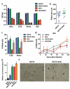
VACV vectors expressing MLKL activate necroptosis-related phenotype in cancer cells. (a) Replication capacity in a panel of cancer cells. Different tumor cell lines were infected with viruses at a MOI of 1 and virus production was measured by plaque-assay 48 hours post-infection. Means +SD are plotted. (b) Plaque size in MA104 cells. MA104 cells were infected at a MOI of 0.05 and, 3 days after infection, the diameter of plaques was measured after dying with crystal violet. MVA vectors were not included in this experiment due to their inability to form plaques. The size of 12 representative plaques and mean ±SD are depicted. (c-d) Cytotoxicity in mouse tumor cell lines. Cells were infected with a MOI of 1 and, 3 days (c) or at different time points after infection (d), % of cells killed was evaluated. (e) Detection of necroptosis-related HMGB1 release. HMGB1 concentration detected by ELISA assay on supernatants of CT26 cells infected with a MOI of 5 with indicated viruses. (f) Images of CT26 cells 24 hours after infection with indicated viruses (MOI of 5). Necroptosis-related swelling of WR/TK-/MLKL-infected cells could be observed (black arrows, scale bar = 25 μm). *, p < .05 vs non-MLKL expressing counterpart.
Figure 3.
Figure 3.
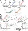
Intratumoral administration of MLKL-expressing vectors protects against primary tumor growth. C57BL/6 mice harboring subcutaneous B16 tumors were randomized and injected twice (days 0 and 4) with an intratumoral dose of 1 × 107 plaque-forming units (pfu) of indicated viruses. Injection of PBS was used as a control. Tumor volume of individual animals (a), mean of treatments (b), and overall survival (c) are plotted for 5–8 mice/group +SD. *, p < .05; **, p < .01; ***, p < .001.
Figure 4.
Figure 4.
VACV-mediated delivery of the necroptosis inducer MLKL activates antitumor immunity. (a-b) Intratumoral administration of MLKL-expressing vectors protects against distal tumors. C57BL/6 mice harboring a primary and a secondary (on the counterflank) B16 tumors were injected twice (days 0 and 4) with an intratumoral dose into the primary tumor of 1 × 107 plaque-forming units (pfu) of indicated viruses. Tumor growth of the untreated secondary tumor of individual animals (a) and mean of treatments (b), tumor growth of the directly injected primary tumor (c), and overall survival (d) are plotted +SD for 5–8 mice/group. (e) Intratumoral administration of MLKL-expressing vectors induces antitumor T cell responses directed against tumor neo-antigens. Mice harboring B16 tumors were treated twice (days 0 and 4) with an intratumoral dose of indicated viruses and, at day 8 after virus-administration, splenocytes were analyzed for their reactivity to indicated peptides by IFN-γ ELISPOT. Individual values of 5 mice/group and mean ±SD are plotted. *, p < .05; **, p < .01; ***, p < .001.

References

    1. Topalian SL, Drake CG, Pardoll DM.. Immune checkpoint blockade: a common denominator approach to cancer therapy. Cancer Cell. 2015. April 13;27(4):450–5. doi:10.1016/j.ccell.2015.03.001. - DOI - PMC - PubMed
    1. O’Donnell JS, Long GV, Scolyer RA, Teng MW, Smyth MJ. Resistance to PD1/PDL1 checkpoint inhibition. Cancer Treat Rev. 2017. January;52:71–81. doi:10.1016/j.ctrv.2016.11.007. - DOI - PubMed
    1. Krysko DV, Garg AD, Kaczmarek A, Krysko O, Agostinis P, Vandenabeele P. Immunogenic cell death and DAMPs in cancer therapy. Nat Rev Cancer. 2012. December;12(12):860–875. doi:10.1038/nrc3380. - DOI - PubMed
    1. Galluzzi L, Buque A, Kepp O, Zitvogel L, Kroemer G. Immunogenic cell death in cancer and infectious disease. Nat Rev Immunol. 2017. February;17(2):97–111. doi:10.1038/nri.2016.107. - DOI - PubMed
    1. Aaes TL, Kaczmarek A, Delvaeye T, De Craene B, De Koker S, Heyndrickx L, Delrue I, Taminau J, Wiernicki B, De Groote P et al. Vaccination with necroptotic cancer cells induces efficient anti-tumor immunity. Cell Rep. 2016. April 12;15(2):274–287. doi:10.1016/j.celrep.2016.03.037. - DOI - PubMed

Publication types