

Primary Pancreatic Large B-Cell Lymphoma Presenting as Acute Pancreatitis
- PMID: 32923189
- PMCID: PMC7478767
- DOI: 10.7759/cureus.9583
Primary Pancreatic Large B-Cell Lymphoma Presenting as Acute Pancreatitis
Abstract
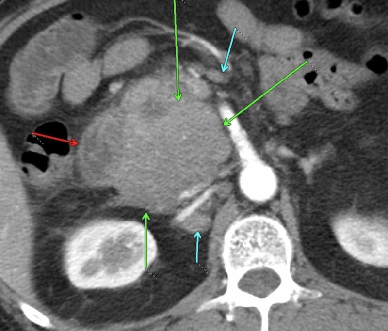
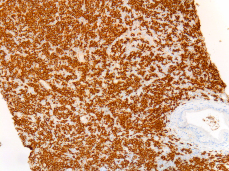
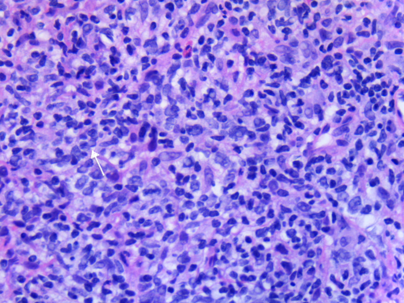
Primary pancreatic lymphoma (PPL) is an extremely rare form of extranodal malignant lymphoma. The most common histological subtype of PPL is diffuse large B-cell lymphoma (DLBCL). Clinical and imaging features of PPL may often overlap with pancreatic adenocarcinoma. Therefore, it is very important to obtain a preoperative cytohistology diagnosis of pancreatic tumors to avoid unnecessary surgeries in cases with a diagnosis of PPL. Herein, we report a 71-year-old male who was admitted to our hospital with a diagnosis of acute pancreatitis after he presented with complaints of nausea, vomiting, and epigastric abdominal pain. MRI of the abdomen revealed a pancreatic head mass, and histopathology and immunohistochemical assessment of the pancreatic lesion established the diagnosis of DLBCL. The patient achieved remission after six cycles of rituximab-cyclophosphamide, doxorubicin (hydroxydaunomycin), vincristine (oncovin), prednisolone (R-CHOP) chemotherapy.
Keywords: acute pancreatitis; diffuse large b cell lymphoma; non-hodgkin lymphoma; primary pancreatic lymphoma.
Copyright © 2020, Tikue et al.
Conflict of interest statement
The authors have declared that no competing interests exist.
Figures
References
-
- Diagnosis and detection of pancreatic cancer. Chu LC, Goggins MG, Fishman EK. Cancer J. 2017;23:333–342. - PubMed
-
- Non-Hodgkin's lymphoma of the gastrointestinal tract: a population-based analysis of incidence, geographic distribution, clinicopathologic presentation features, and prognosis. Danish Lymphoma Study Group. d'Amore F, Brincker H, Grønbaek K, et al. J Clin Oncol. 1994;12:1673–1684. - PubMed
-
- Occurrence and prognosis of extranodal lymphomas. Freeman C, Berg JW, Cutler SJ. https://pubmed.ncbi.nlm.nih.gov/5007387/ Cancer. 1972;29:252–260. - PubMed
Publication types
LinkOut - more resources
Full Text Sources
Research Materials