. 2020 Aug 13:10:1410.
doi: 10.3389/fonc.2020.01410. eCollection 2020.

Machine Learning-Based Radiomics Nomogram Using Magnetic Resonance Images for Prediction of Neoadjuvant Chemotherapy Efficacy in Breast Cancer Patients

Affiliations

Machine Learning-Based Radiomics Nomogram Using Magnetic Resonance Images for Prediction of Neoadjuvant Chemotherapy Efficacy in Breast Cancer Patients

Shujun Chen et al. Front Oncol. .

Abstract

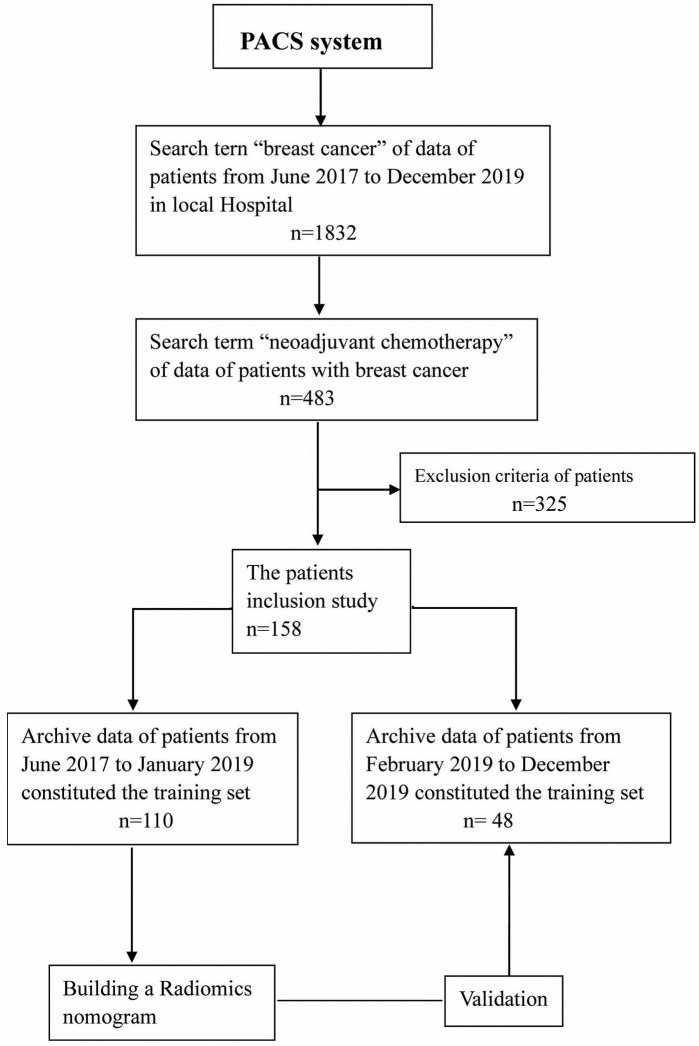
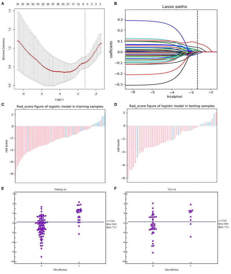
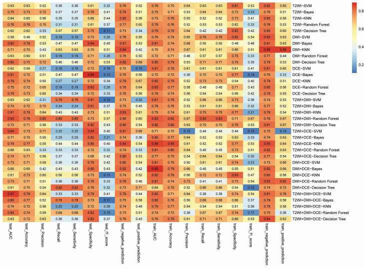
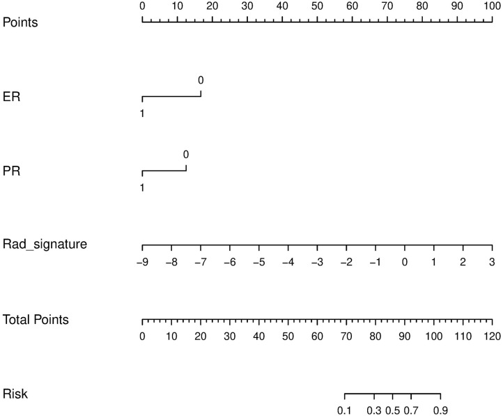
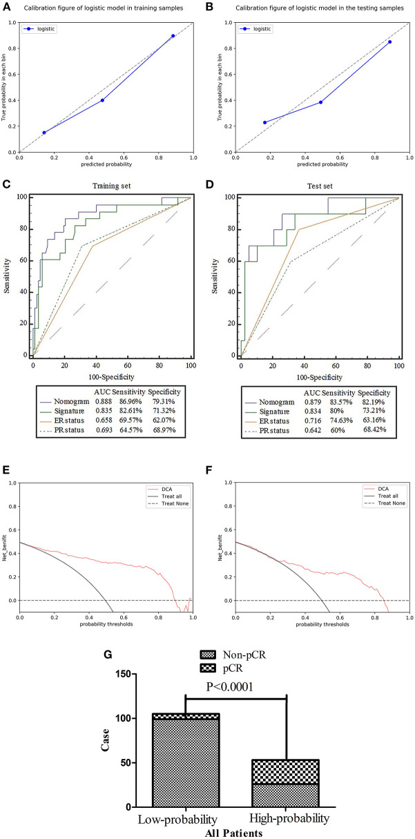
Purpose: The construction and validation of a radiomics nomogram based on machine learning using magnetic resonance image (MRI) for predicting the efficacy of neoadjuvant chemotherapy (NACT) in patients with breast cancer (BCa). Methods: This retrospective investigation consisted of 158 patients who were diagnosed with BCa and underwent MRI before NACT, of which 33 patients experienced pathological complete response (pCR) by the postoperative pathological examination. The patients with BCa were divided into the training set (n = 110) and test set (n = 48) randomly. The features were selected by the maximum relevance minimum redundancy (mRMR) and absolute shrinkage and selection operator (LASSO) algorithm in the training set. In return, the radiomics signature was established using machine learning. The predictive score of each patient was calculated using the radiomics signature formula. Finally, the predictive scores and clinical factors were used to perform the multivariate logistic regression and construct the nomogram. Receiver operating characteristics (ROC) analyses were used to assess and validate the diagnostic accuracy of the nomogram in the test set. Lastly, the usefulness of the nomogram was confirmed via decision curve analysis (DCA). Results: The radiomics signature was well-discriminated in the training set [AUC 0.835, specificity 71.32%, and sensitivity 82.61%], and test set (AUC 0.834, specificity 73.21%, and sensitivity 80%). Containing the radiomics signature and hormone status, the radiomics nomogram showed good calibration and discrimination in the training set [AUC 0.888, specificity 79.31%, and sensitivity 86.96%] and test set (AUC 0.879, specificity 82.19%, and sensitivity 83.57%). The decision curve indicated the clinical usefulness of our nomogram. Conclusion: Our radiomics nomogram showed good discrimination in patients with BCa who experience pCR after NACT. The model may aid physicians in predicting how specific patients may respond to BCa treatments in the future.

Keywords: breast cancer; machine learning; neoadjuvant chemotherapy; nomogram; pathological complete response; radiomics.


Figures

Figure 1
Figure 1
Flowchart showing the recruitment of patients and the overall design of this retrospective study.
Figure 2
Figure 2
Workflow for building the radiomics signature and creating the model. T2WI, T2-weighted image; DWI, diffusion-weighted imaging; DCE, dynamic contrast enhancement.
Figure 3
Figure 3
Texture feature selection using the LASSO logistic regression. (A) The tuning parameter (λ) selection in the LASSO model used 10-fold cross-validation via the minimum criteria. Partial likelihood deviance was plotted vs. log (λ). The dotted vertical lines were drawn at the optimal values using the minimum criteria and the 1-SE criteria. (B) The LASSO coefficient profiles of the 35 texture features. The vertical line was drawn at the value selected using 10-fold cross-validation in the log (λ) sequence, and six features with non-zero coefficients are indicated. Score diagrams of the radiomics signature in the (C) training set and (D) test set. Red represents non-pCR and blue represents pCR. A score >0 indicates pCR, and a score <0 indicates non-pCR. Both panels (C,D) show interactive dot diagrams revealing the accuracy of the radiomics signature for predicting pCR in patients of the (E) training set and (F) test set. Zero represents non-pCR, and 1 represents pCR. The horizontal line indicates the best threshold point to distinguish patients with pCR from patients with non-pCR.
Figure 4
Figure 4
The evaluation results of radiomics signature combined features from different imaging sequences and machine learning methods. The right column of the figure shows the combination of different sequences. The horizontal coordinate shows different items to be evaluated in the training set and the test set, and the value in each frame represents the evaluated result of the items of the corresponding sequence combination. The closer the color is to red, the greater the value. In this study, the larger the AUC value represents the better combined model built by machine learning.
Figure 5
Figure 5
Radiomics nomogram to predict the patient with pCR. The radiomics nomogram was developed in the training set, with the rad-score, ER status, and PR status.
Figure 6
Figure 6
(A,B) Calibration of the radiomics nomogram for predicting pCR in the training and test sets. Dashed line is the reference line where an ideal nomogram would lie, while the solid line corrects for any bias in the hybrid nomogram. (C,D) Evaluation of the accuracy of the ER status, PR status, radiomics signature, and nomogram for predicting pCR in the training and test sets. (E,F) Decision curve analysis was used to show the clinical effect of the nomogram in the prediction of pCR in breast cancer (BCa) patients in training and test sets. (G) Probability of pCR in the high-probability group was significantly higher than that in the low-probability group (p < 0.0001).

References

    1. Siegel RL, Miller KD, Jemal A. Cancer statistics 2016. CA Cancer J Clin. (2016) 66:7–30. 10.3322/caac.21332 - DOI - PubMed
    1. Early Breast Cancer Trialists' Collaborative Group (EBCTCG) . Long-term outcomes for neoadjuvant versus adjuvant chemotherapy in early breast cancer: meta-analysis of individual patient data from ten randomised trials. Lancet Oncol. (2018) 19:27–39. 10.1016/S1470-2045(17)30777-5 - DOI - PMC - PubMed
    1. Barroso-Sousa R, Silva DD, Alessi JV, Mano MS. Neoadjuvant endocrine therapy in breast cancer: current role and future perspectives. Ecancermedicalscience. (2016) 10:609. 10.3332/ecancer.2016.609 - DOI - PMC - PubMed
    1. Untch M, Konecny GE, Paepke S, von Minckwitz G. Current and future role of neoadjuvant therapy for breast cancer. Breast. (2014) 23:526–537. 10.1016/j.breast.2014.06.004 - DOI - PubMed
    1. Rastogi P, Anderson SJ, Bear HD, Geyer CE, Kahlenberg MS, Robidoux A, et al. . Preoperative chemotherapy: updates of National Surgical Adjuvant Breast and Bowel Project Protocols B-18 and B-27. J Clin Oncol. (2008) 26:778–85. 10.1200/JCO.2007.15.0235 - DOI - PubMed

LinkOut - more resources