. 2020 Aug 28;6(35):eaaz0127.
doi: 10.1126/sciadv.aaz0127. eCollection 2020 Aug.

Ingestible transiently anchoring electronics for microstimulation and conductive signaling

Affiliations

Ingestible transiently anchoring electronics for microstimulation and conductive signaling

Alex Abramson et al. Sci Adv. .

Abstract

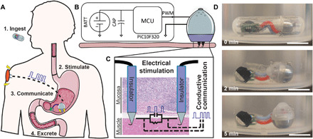
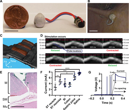
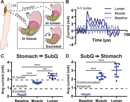
Ingestible electronic devices enable noninvasive evaluation and diagnosis of pathologies in the gastrointestinal (GI) tract but generally cannot therapeutically interact with the tissue wall. Here, we report the development of an orally administered electrical stimulation device characterized in ex vivo human tissue and in in vivo swine models, which transiently anchored itself to the stomach by autonomously inserting electrically conductive, hooked probes. The probes provided stimulation to the tissue via timed electrical pulses that could be used as a treatment for gastric motility disorders. To demonstrate interaction with stomach muscle tissue, we used the electrical stimulation to induce acute muscular contractions. Pulses conductively signaled the probes' successful anchoring and detachment events to a parenterally placed device. The ability to anchor into and electrically interact with targeted GI tissues controlled by the enteric nervous system introduces opportunities to treat a multitude of associated pathologies.


Figures

Fig. 1
Fig. 1. Self-orienting technology for injection and electrical microstimulation (STIMS).
(A) STIMS injects electrically conductive hooked needles into the tissue for retention and uses them to stimulate the tissue via timed electrical pulses. The pulses also allow the device to communicate conductively to the subcutaneous space of the animal and convey proper device attachment and excretion. (B) STIMS architecture. BATT, Battery; CAP, capacitor; MCU, microcontroller unit; PWM, pulse width modulation. (C) Only the tips of the probes are conductive, which allows the electrical pulses to target a specific layer of tissue. Photo credit: Alex Abramson, MIT. (D) STIMS fits inside a 000 capsule, which is 26.1 mm in length and 9.9 mm in diameter, to aid in ingestion. The device includes a self-orienting system and autonomous injection mechanism, a microcontroller for pulse generation, and two coin batteries for power. A STIMS capsule was placed in 35° to 45°C water under agitation, and the device self-oriented after being released. Scale bars, 1 cm. Photo credit: Alex Abramson, MIT.
Fig. 2
Fig. 2. Implanted hooked needles generate retention forces that allow devices to remain localized to the stomach wall for prolonged time periods.
(A) Scolex of the Taenia solium tapeworm, a parasitic worm that uses hooks to attach to the GI tract of its host. For scale, the diameter of a typical scolex for this species is 1 mm. Photo credit: Centers of Disease Control, public domain. (B) Hooks at the end of a 32-gauge needle as small as 30 μm in size latched onto ex vivo human stomach tissue. Scale bar, 5 mm. Photo credit: Alex Abramson, MIT. (C) Hooked needles fabricated in different sizes and combined into arrays created an attachment mechanism similar to the tapeworm, Scale bar, 100 μm. Photo credit: Alex Abramson, MIT. (D) The STIMS device inserted arrays of hooked needles into the stomach tissue. A 3D CAD model of the STIMS device, Scale bar, 5 mm. (E) Pullback forces from hooked needles inserted into ex vivo swine stomach (n = 3). (F) Pullback forces from hooked needles inserted into swine and human stomach tissues (n = 5). “No hook” data represent frictional pullback force from ex vivo swine tissue when using needles that do not hook onto the tissue. (G) The relative anchoring force was linearly correlated with the number of needles inserted into tissue (n = 9 over three stomachs). (H) Increasing the distance between the inserted needles increased the anchoring force (n = 5). (I) Anchoring forces were on average 0.7 N in both in vivo and ex vivo swine stomachs when using a STIMS device with three needles spaced 1 mm apart (n = 6 over two stomachs). Means ± SD; *P < 0.05; **P < 0.01; ***P < 0.001.
Fig. 3
Fig. 3. Needle insertion force profiles for human and swine stomachs.
(A) Penetration forces in the body, fundus, and antrum of human stomachs (n = 9 over three stomachs) compared with penetration forces in the body of ex vivo swine stomachs (n = 12 over three stomachs). (B) Swine in vivo penetration forces with multiple sizes of needles (n = 15 over three stomach). (C) Ex vivo and in vivo perforation experiments in swine stomachs (n = 15 over three stomachs) showing the force and (D) displacement required by a needle coated in tissue marking dye to penetrate dye completely through the mucosa and to completely perforate through the muscular tissue. Means ± SD; ***P < 0.001; ****P < 0.0001.
Fig. 4
Fig. 4. In vivo electrical stimulation of the gut.
(A) STIMS system connected to two 1.55-V silver oxide coin cell batteries and a microcontroller. Before ingestion, electronics were coated in PDMS. Photo credit: Alex Abramson, MIT. (B) In vivo release and tissue localization of the STIMS. Photo credit: Joy Collins, MIT. (C) Stacked 2D and (D) sequential ultrasound images over time of muscular stimulation from electrical probes in in vivo swine stomachs. In (C), artificially colored red areas denote contraction events. MATLAB was used to isolate the muscle section from the rest of the image. A video is included in the supplement. Photo credit: Joy Collins, MIT. (E) Histology from ex vivo swine tissue injected by STIMS with hooked needles using a 5-N spring. Needles inserted through the mucosal layer of the stomach without perforating the tissue (M, mucosa; SM, submucosa; Mu, outer muscle). Photo credit: Kathleen Cormier, MIT. (F) Maximum current measured through a circuit connected in series with an in vivo swine stomach using a 2.5-V microcontroller (n = 5). ST, stomach; SI, small intestine. (G) Microcontroller stimulation events in vivo measured via an oscilloscope (n = 15). Scale bars, 1 cm (A and B) and 1 mm (E). Means ± SD; *P < 0.05; **P < 0.01; ***P < 0.001; ****P < 0.0001.
Fig. 5
Fig. 5. In vivo conductive communication conveys tissue wall localization.
(A) The conductive nature of the body allows electrical pulses to pass through the tissue from the stomach to the subcutaneous space. (B) Current readout from a circuit branch containing a 10-ohm resistor connected to the subcutaneous space of a swine when a 330-μs, 5-V electrical pulse is generated by a microcontroller in the stomach. Electrical pulses have different profiles depending on if they originate in the stomach lumen or stomach muscle. (C) Average current of the pulse passing through the body in the subcutaneous space when a pulse is generated in the stomach. Depending on the current reading, it is possible to discern whether probes are in the stomach muscle, are floating in GI luminal fluid, or are not in contact with the body. (D) Pulses generated in the subcutaneous space can be read in the stomach as well. Means ± SD; ***P < 0.001; ****P < 0.0001.

References

    1. Kaelberer M. M., Buchanan K. L., Klein M. E., Barth B. B., Montoya M. M., Shen X., Bohórquez D. V., A gut-brain neural circuit for nutrient sensory transduction. Science 361, eaat5236 (2018). - PMC - PubMed
    1. Han W., Tellez L. A., Perkins M. H., Perez I. O., Qu T., Ferreira J., Ferreira T. L., Quinn D., Liu Z. W., Gao X. B., Kaelberer M. M., Bohórquez D. V., Shammah-Lagnado S. J., de Lartigue G., de Araujo I. E., A neural circuit for gut-induced reward. Cell 175, 665–678.e23 (2018). - PMC - PubMed
    1. Abell T., McCallum R., Hocking M., Koch K., Abrahamsson H., LeBlanc I., Lindberg G., Konturek J., Nowak T., Quigley E. M. M., Tougas G., Starkebaum W., Gastric electrical stimulation for medically refractory gastroparesis. Gastroenterology 125, 421–428 (2003). - PubMed
    1. Rao M., Gershon M. D., The bowel and beyond: The enteric nervous system in neurological disorders. Nat. Rev. Gastroenterol. Hepatol. 13, 517–528 (2016). - PMC - PubMed
    1. Sampson T. R., Mazmanian S. K., Control of brain development, function, and behavior by the microbiome. Cell Host Microbe 17, 565–576 (2015). - PMC - PubMed

Publication types

LinkOut - more resources