. 2020 Sep 1:4:24.
doi: 10.1038/s41698-020-00129-0. eCollection 2020.

Honeybee venom and melittin suppress growth factor receptor activation in HER2-enriched and triple-negative breast cancer

Affiliations

Honeybee venom and melittin suppress growth factor receptor activation in HER2-enriched and triple-negative breast cancer

Ciara Duffy et al. NPJ Precis Oncol. .

Abstract

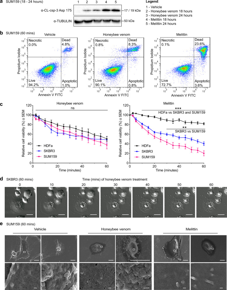
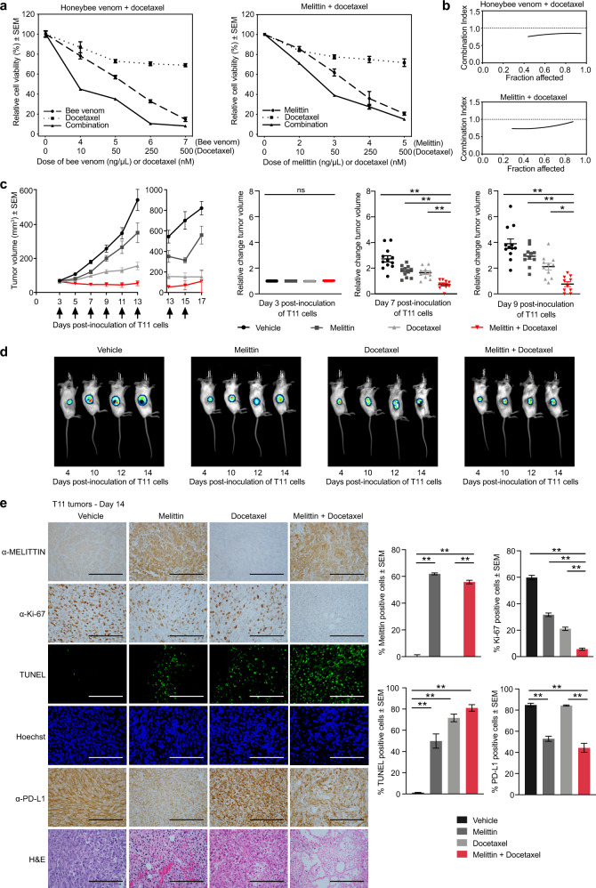
Despite decades of study, the molecular mechanisms and selectivity of the biomolecular components of honeybee (Apis mellifera) venom as anticancer agents remain largely unknown. Here, we demonstrate that honeybee venom and its major component melittin potently induce cell death, particularly in the aggressive triple-negative and HER2-enriched breast cancer subtypes. Honeybee venom and melittin suppress the activation of EGFR and HER2 by interfering with the phosphorylation of these receptors in the plasma membrane of breast carcinoma cells. Mutational studies reveal that a positively charged C-terminal melittin sequence mediates plasma membrane interaction and anticancer activity. Engineering of an RGD motif further enhances targeting of melittin to malignant cells with minimal toxicity to normal cells. Lastly, administration of melittin enhances the effect of docetaxel in suppressing breast tumor growth in an allograft model. Our work unveils a molecular mechanism underpinning the anticancer selectivity of melittin, and outlines treatment strategies to target aggressive breast cancers.

Keywords: Breast cancer; Molecular medicine.


Conflict of interest statement

Competing interestsK.P. received funding from Promega, BMG Labtech, and Dimerix as ARC Linkage Grant participating organizations. These participating organizations played no role in any aspect of the conception or design of the research, collection, analysis and interpretation of the results, or writing and editing of the paper. K.P. is the chief scientific advisor of Dimerix, of which he maintains a shareholding. Remaining authors declare no competing interests.

Figures

Fig. 1
Fig. 1. Honeybee venom and melittin specifically reduce breast tumor cell viability.
a The process of bee venom collection and melittin treatment of breast cancer cells, featuring a honeybee collected in Australia. b Cell-viability assays of a panel of human normal and breast cancer cell lines treated with honeybee venom from Australia (left) or melittin (right), with c the IC50 values (generalized linear models). Cell-viability assays of normal human dermal fibroblasts (HDFa) and breast cancer cell lines (SUM159 and SKBR3) treated with d venom from populations of honeybees in Ireland (left) and England (right) (one-way ANOVA), and e venom from England worker (left) and queen (right) bumblebees. f Absorbance (405 nm) of aqueous solutions of melittin and bee venom assessed by ELISA with the anti-melittin antibody and IgG control (two-way ANOVA). g Cell-viability assays in HDFa and SUM159 cells after blocking melittin using the anti-melittin antibody with honeybee venom (left) and melittin (right). Data are represented as mean ± SEM (n = 3). Differences were considered significant at p < 0.05 (*), p < 0.01 (**), and p < 0.001 (***). See also Supplementary Fig. 1.
Fig. 2
Fig. 2. Honeybee venom and melittin induce apoptosis and membrane disruption.
a Western blot for the detection of cleaved caspase-3 (CL-csp-3) in SUM159 cells treated with vehicle (1), honeybee venom (2–3), and melittin (4–5) for 18 and 24 h. b Flow cytometry analysis of SUM159 cells treated with the IC50 of honeybee venom (5.58 ng/µL) and the IC50 of melittin (4.24 ng/µL) for 1 h. c Cell-viability temporal response assays of normal human dermal fibroblasts (HDFa) and breast cancer cells (SUM159 and SKBR3) treated with honeybee venom (left) or melittin (right) over 1 hour (two-way ANOVAs). d Live-cell confocal microscopy of SKBR3 cells treated with the IC50 of honeybee venom (5.77 ng/µL) over 1 h, with time in minutes post treatment. Scale bars represent 15 µm. e Scanning electron microscopy of SUM159 cells treated with the IC50 of honeybee venom (5.58 ng/µL) and the IC50 of melittin (4.24 ng/µL) over 1 h, with two representative images shown for each treatment group. The white outline in the top images indicates the respective regions of each cell in the bottom images. Scale bars represent 10 µm (top row) and 200 nm (bottom row). Data are represented as mean ± SEM (n = 3). Differences were considered significant at p < 0.05 (*), p < 0.01 (**), and p < 0.001 (***). See also Supplementary Figs. 2, 10, and 16.
Fig. 3
Fig. 3. Engineering melittin with an RGD motif enhances breast cancer selectivity.
a Cell-viability assays of TNBC (SUM159) and HER2-enriched breast cancer (SKBR3) cells treated with DEDE-melittin for 24 h. b Cell-viability assays of T11 cells treated with melittin, RGD1-melittin, SV40-melittin, and DEDE-melittin for 24 h (t test). c Cell-viability assays of normal human dermal fibroblasts (HDFa) and SUM159 treated with melittin (left) and RGD1-melittin (right) for 24 h (t tests). d Western blot for the detection of cleaved caspase-3 (CL-csp-3) in lysates from SUM159 cells treated with vehicle, melittin, DEDE-melittin, or RGD1-melittin for 24 h. e Absorbance (405 nm) of aqueous solutions of melittin, RGD1-melittin, DEDE-melittin, and SV40-melittin subjected to an ELISA with the anti-melittin antibody (two-way ANOVA). f The amino-acid sequence and top predicted 3D model of melittin (green), RGD1-melittin (purple), DEDE-melittin (blue), and SV40-melittin (orange). g Immunofluorescence images of SUM159 treated with vehicle, honeybee venom, melittin, RGD1-melittin, or DEDE-melittin for 30 min. In blue: cell nuclei, in red: anti-EGFR, and in green: anti-melittin. The white outlines in the merged images indicate the respective regions in the zoomed images. Scale bars represent 25 µm, and 6.25 µm for the zoomed images. Data are represented as mean ± SEM (n = 3). Differences were considered significant at p < 0.05 (*), p < 0.01 (**), and p < 0.001 (***). See also Supplementary Figs. 3 and 10.
Fig. 4
Fig. 4. Honeybee venom and melittin suppress the phosphorylation of EGFR and HER2.
a Phosphorylation kinetics of HER2, EGFR, and downstream MAPK and Akt pathways after treatment with honeybee venom and melittin in SKBR3 (left) and SUM159 (right) breast cancer cells, assessed by immunoblotting. b Bioluminescence resonance energy transfer (BRET) kinetic analysis of TAMRA-EGF, FITC-melittin, and FITC–DEDE-melittin interaction with NanoLuc-EGFR in HEK293FT cells. The peptides were added after the cells were equilibrated in the reader with the NanoLuc substrate furimazine for 5 min. c Saturation-binding analysis of increasing concentrations of TAMRA-EGF, FITC-melittin, and FITC–DEDE-melittin in HEK293FT cells transfected with NanoLuc-EGFR in the presence or absence of unlabeled EGF (1 µM). Data are expressed as raw BRET ratios and represented as mean ± SEM (n = 3, two-way ANOVA). d Proposed model of action of melittin interfering with the dimerization and phosphorylation of RTKs in the plasma membrane. Differences were considered significant at p < 0.05 (*), p < 0.01 (**), and p < 0.001 (***). See also Supplementary Figs. 4–6 and 11–15.
Fig. 5
Fig. 5. Melittin sensitizes highly aggressive TNBC tumors to docetaxel treatment in vivo.
a Cell-viability assays of T11 cells treated with honeybee venom and melittin alone and in combination with docetaxel for 24 h. Representative plots of the combination treatments are presented (n = 3). b Combination index graphs obtained for different fractions of cells affected in each combination, calculated using CompuSyn software. c Tumor volumes of mice treated intratumorally with vehicle, 5 mg/kg melittin, 7 mg/kg docetaxel, and 5 mg/kg melittin + 7 mg/kg docetaxel. Arrows indicate the treatment days. Corresponding scatter plots of relative change in tumor volumes at days 3, 7, and 9 are indicated (one-way ANOVA, n = 12). d Representative bioluminescence imaging (BLI) of T11-luciferase tumors in mice at days 4, 10, 12, and 14 post inoculation of the cells. e Representative images of immunohistochemistry and immunofluorescence in tumor biopsies from mice extracted on day 14 post T11 inoculation stained with anti-melittin, anti-Ki-67, TUNEL assay, Hoechst, anti-PD-L1, and H&E (one-way ANOVA, n = 8). Scale bars represent 100 µm. Data are represented as mean ± SEM. Differences were considered significant at p < 0.05 (*), p < 0.01 (**), and p < 0.001 (***). See also Supplementary Figs. 7–9.

References

    1. Son DJ, et al. Therapeutic application of anti-arthritis, pain-releasing, and anti-cancer effects of bee venom and its constituent compounds. Pharmacol. Ther. 2007;115:246–270. - PubMed
    1. Fitzmaurice C, et al. Global, regional, and national cancer incidence, mortality, years of life lost, years lived with disability, and disability-adjusted life-years for 32 Cancer Groups, 1990 to 2015. JAMA Oncol. 2017;3:524. - PMC - PubMed
    1. Park S-C, et al. Investigation of toroidal pore and oligomerization by melittin using transmission electron microscopy. Biochem. Biophys. Res. Commun. 2006;343:222–228. - PubMed
    1. Lyu Y, Zhu X, Xiang N, Narsimhan G. Molecular Dynamics Study of Pore Formation by Melittin in a 1,2-Dioleoyl- sn -glycero-3-phosphocholine and 1,2-Di(9 Z -octadecenoyl)- sn -glycero-3-phospho-(1′- rac -glycerol) Mixed Lipid Bilayer. Ind. Eng. Chem. Res. 2015;54:10275–10283.
    1. Terwilliger TC, Eisenberg D. The structure of melittin. II. Interpretation of the structure. J. Biol. Chem. 1982;257:6016–6022. - PubMed