Review
. 2019 Mar 11;1(1):H1-H8.
doi: 10.1530/VB-19-0008. eCollection 2019.

Metabolic regulation of exercise-induced angiogenesis

Affiliations
Review

Metabolic regulation of exercise-induced angiogenesis

Tatiane Gorski et al. Vasc Biol. .

Abstract

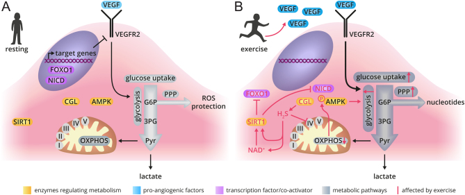
Skeletal muscle relies on an ingenious network of blood vessels, which ensures optimal oxygen and nutrient supply. An increase in muscle vascularization is an early adaptive event to exercise training, but the cellular and molecular mechanisms underlying exercise-induced blood vessel formation are not completely clear. In this review, we provide a concise overview on how exercise-induced alterations in muscle metabolism can evoke metabolic changes in endothelial cells (ECs) that drive muscle angiogenesis. In skeletal muscle, angiogenesis can occur via sprouting and splitting angiogenesis and is dependent on vascular endothelial growth factor (VEGF) signaling. In the resting muscle, VEGF levels are controlled by the estrogen-related receptor γ (ERRγ). Upon exercise, the transcriptional coactivator peroxisome-proliferator-activated receptor-γ coactivator-1α (PGC1α) orchestrates several adaptations to endurance exercise within muscle fibers and simultaneously promotes transcriptional activation of Vegf expression and increased muscle capillary density. While ECs are highly glycolytic and change their metabolism during sprouting angiogenesis in development and disease, a similar role for EC metabolism in exercise-induced angiogenesis in skeletal muscle remains to be elucidated. Nonetheless, recent studies have illustrated the importance of endothelial hydrogen sulfide and sirtuin 1 (SIRT1) activity for exercise-induced angiogenesis, suggesting that EC metabolic reprogramming may be fundamental in this process. We hypothesize that the exercise-induced angiogenic response can also be modulated by metabolic crosstalk between muscle and the endothelium. Defining the underlying molecular mechanisms responsible for skeletal muscle angiogenesis in response to exercise will yield valuable insight into metabolic regulation as well as the determinants of exercise performance.

Keywords: angiogenesis; endothelial metabolism; exercise; metabolism; microvasculature.


Conflict of interest statement

The authors declare that there is no conflict of interest that could be perceived as prejudicing the impartiality of this review.

Figures

Figure 1
Figure 1
Exercise-induced activation of transcription factors in myofibers stimulates angiogenesis. While ERRγ determines baseline muscle vascularization either via direct binding to the VEGF promoter or via controlling the activity of AMPK, exercise-induced activation of PGC1α (through recruitment of ERRα to the VEGF promotor), and potentially increased stabilization of HIF1α, culminates in the increased expression of VEGF and other pro-angiogenic factors. Release of VEGF and other angiogenic factors from the exercising muscle leads to increased muscle vascularization through vessel sprouting or vessel splitting. AMPK, 5′ adenosine monophosphate-activated protein kinase; ERRα, estrogen-related receptor α; ERRγ, estrogen-related receptor γ; HIF1α, hypoxia inducible factor 1α; PGC1α, peroxisome-proliferator-activated receptor-γ coactivator-1α; VEGF, vascular endothelial growth factor.
Figure 2
Figure 2
Proposed model for exercise-induced changes in EC metabolism promoting angiogenesis. (Panel A) ECs are highly glycolytic and produce 85% of their energy glycolytically. Mitochondria do not significantly contribute to energy production but rather maintain NAD+/NADH balance. (Panel B) During exercise, increased CGL activity leads to H2S generation. CGL-derived H2S downregulates endothelial OXPHOS by inhibiting complex IV activity. This leads to increased glucose uptake, glycolysis and pentose phosphate pathway flux in an AMPK-dependent fashion. In addition, exercise-induced angiogenesis requires adequate activation of SIRT1, at least partially because endothelial SIRT1 is required for VEGF-mediated angiogenesis. SIRT1 also controls EC function by inhibiting Notch (NICD indicates active Notch) and via inactivating FOXO1 (forkhead box O1, not shown). 3PG, glyceraldehyde 3-phosphate; AMPK, 5′ adenosine monophosphate-activated protein kinase; CGL, cystathionine γ-lyase; G6P, glucose 6-phosphate; NAD, nicotinamide adenine dinucleotide; NICD, Notch intracellular domain; OXPHOS, oxidative phosphorylation; PPP, pentose phosphate pathway; Pyr, pyruvate; ROS, reactive oxygen species; SIRT1, Sirtuin 1; VEGF, vascular endothelial growth factor; VEGFR2, vascular endothelial growth factor receptor 2.

References

    1. DeFronzo RA, Jacot E, Jequier E, Maeder E, Wahren J, Felber JP. The effect of insulin on the disposal of intravenous glucose. Results from indirect calorimetry and hepatic and femoral venous catheterization. Diabetes 1981. 30 1000–1007. (10.1016/j.cja.2015.03.007) - DOI - PubMed
    1. Meyer C, Dostou JM, Welle SL, Gerich JE. Role of human liver, kidney, and skeletal muscle in postprandial glucose homeostasis. American Journal of Physiology: Endocrinology and Metabolism 2002. 282 E419–E427. (10.1152/ajpendo.00032.2001) - DOI - PubMed
    1. Ahlborg G, Felig P, Hagenfeldt L, Hendler R, Wahren J. Substrate turnover during prolonged exercise in man. Journal of Clinical Investigation 1974. 53 1080–1090. (10.1172/JCI107645) - DOI - PMC - PubMed
    1. Egan B, Zierath JR. Exercise metabolism and the molecular regulation of skeletal muscle adaptation. Cell Metabolism 2013. 17 162–184. (10.1016/j.cmet.2012.12.012) - DOI - PubMed
    1. Murrant CL, Sarelius IH. Coupling of muscle metabolism and muscle blood flow in capillary units during contraction. Acta Physiologica Scandinavica 2000. 168 531–541. (10.1046/j.1365-201X.2000.00706.x) - DOI - PubMed

LinkOut - more resources