Review
. 2020 Mar 20;2(1):H1-H18.
doi: 10.1530/VB-19-0033. eCollection 2020.

Immune cell trafficking across the blood-brain barrier in the absence and presence of neuroinflammation

Affiliations
Review

Immune cell trafficking across the blood-brain barrier in the absence and presence of neuroinflammation

Luca Marchetti et al. Vasc Biol. .

Abstract

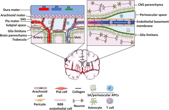
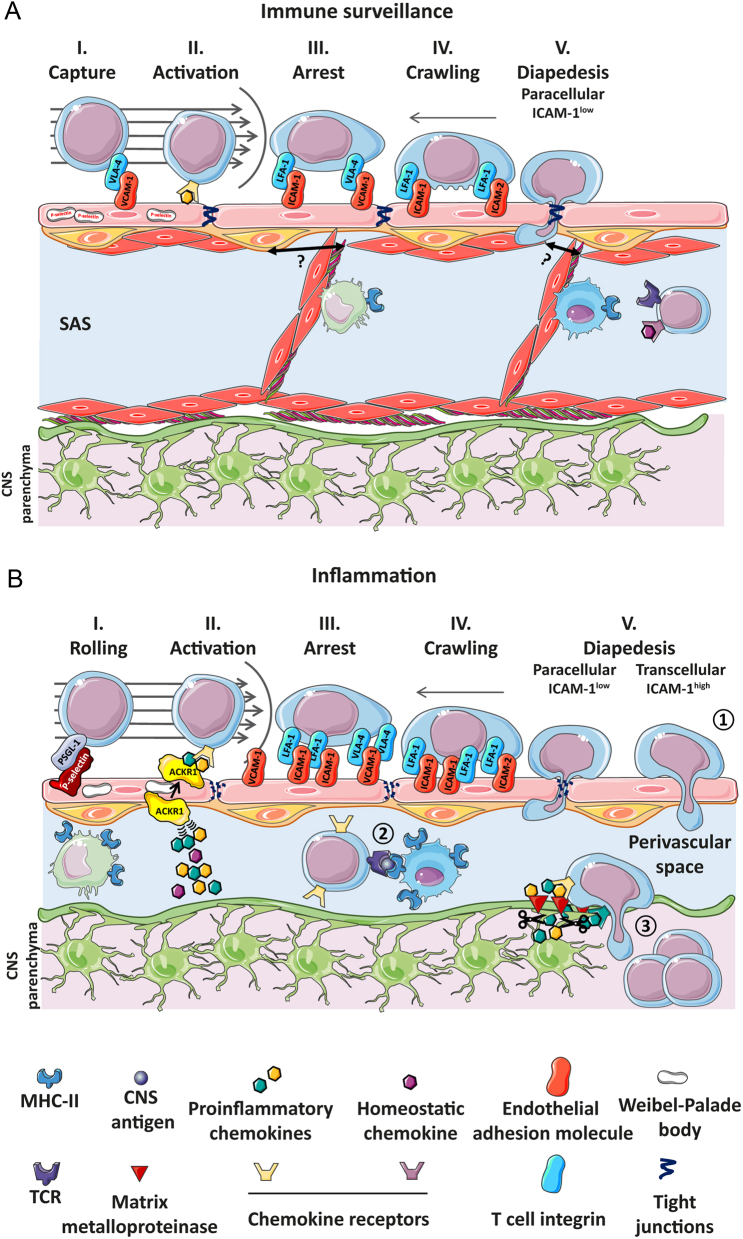
To maintain the homeostatic environment required for proper function of CNS neurons the endothelial cells of CNS microvessels tightly regulate the movement of ions and molecules between the blood and the CNS. The unique properties of these blood vascular endothelial cells are termed blood-brain barrier (BBB) and extend to regulating immune cell trafficking into the immune privileged CNS during health and disease. In general, extravasation of circulating immune cells is a multi-step process regulated by the sequential interaction of adhesion and signalling molecules between the endothelial cells and the immune cells. Accounting for the unique barrier properties of CNS microvessels, immune cell migration across the BBB is distinct and characterized by several adaptations. Here we describe the mechanisms that regulate immune cell trafficking across the BBB during immune surveillance and neuroinflammation, with a focus on the current state-of-the-art in vitro and in vivo imaging observations.

Keywords: blood-brain barrier; immune cell migration; life cell; multiple sclerosis; neuroinflammation.


Conflict of interest statement

The authors declare that there is no conflict of interest that could be perceived as prejudicing the impartiality of the research reported.

Figures

Figure 1
Figure 1
Leptomeningeal and parenchymal blood-brain barrier. The meninges at the surface of the brain (left) are composed by three layers, namely the dura mater, the arachnoid mater and the pia mater. In the dura mater we find dural arteries (DA) and veins (DV), as well as dural lymphatic vessels (DL). Dural blood vessels do not form a blood-brain barrier. The cells of the arachnoid mater form a blood-cerebrospinal fluid barrier (BCSFB) between the dura mater and the cerebrospinal fluid (CSF)-filled subarachnoid space (SAS). In humans the arachnoid mater is composed of several layers of arachnoid cells. The SAS harbors antigen-presenting cells (APCs), i.e. subarachnoid macrophages. Blood vessels in the SAS are ensheathed by a layer of pia mater, further connected to the arachnoid mater by trabeculae spanning the SAS. The center of the trabeculae is composed of a collagen core that is covered by cells of the pia mater. A thin layer of pia mater also covers the arteries that dive into the brain. The glia limitans is composed of the parenchymal basement membrane and astrocyte foot-processes and covers as glia limitans superficialis the entire surface of the CNS parenchyma and accompanies as glia limitans perivascularis the blood vessels in the CNS. Venules in the SAS and subpial space form a BBB albeit they lack ensheathment by astrocyte endfeet. The arachnoid and pia maters are referred to as leptomeninges. The anatomical details have been summarized in (5). The BBB at the level of CNS parenchymal vessels (right inset) is composed by highly specialized endothelial cells, held together by molecularly unique and complex tight junction strands. Pericytes are embedded in the endothelial basement membrane, while the glia limitans further ensheaths the CNS microvasculature. At the level of the capillaries, the endothelial basement membrane and glia limitans are fused. At the postcapillary venules, where immune cell trafficking takes place, the two basement membranes are separated by the CSF-filled perivascular space, which harbors rare antigen-presenting cells. Drawings of the individual cell types were adapted from Servier Medical Art (http://smart.servier.com/), licensed under a Creative Common Attribution 3.0 Generic License.
Figure 2
Figure 2
Multi-step T-cell extravasation across the BBB during heath and neuroinflammation. T-cell extravasation across subarachnoid venules during immune surveillance (A) or across BBB postcapillary venules during inflammation (B) is depicted. Leptomeningeal endothelial cells store P-selectin in Weibel-Palade bodies, however, in the absence of inflammation α4β1-mediated capture is the most observed first interaction. After GPCR-mediated shear-Figure 2 Continuedresistant arrest, T cells crawl against the direction of the flow and cross the BBB endothelium preferentially via the paracellular pathway. Pial cells are reported to partially cover the venular wall in the SAS (highlighted by the question mark), but do not seem to establish a barrier for T cell extravasation. In the absence of CNS antigens presented by subarachnoid macrophages and dendric cells on MHC-II molecules, T cells will not cross the glia limitans and may rather be flushed away with the CSF. During inflammation, leptomeningeal but also parenchymal BBB endothelial cells (B) allow for activated T-cell rolling, mediated by P-selectin which is de novo expressed as it is not stored in Weibel-Palade bodies. Inflammatory chemokines produced by astrocytes are transported from the abluminal to the luminal side of the BBB by ACKR1. After their GPCR-dependent arrest, T cells crawl on endothelial ICAM-1 and ICAM-2 against the direction of the flow with increased levels of endothelial ICAM-1 leading to increased transcellular T cell diapedesis. Once T cells have crossed the BBB endothelium (1), CNS-antigen-specific T cells may recognize their cognate antigens on perivascular APCs (2) and become reactivated behind the BBB. Matrix metalloproteinases produced by infiltrating and perivascular-activated myeloid cells as well as astrocytes cleave the astrocytic endfeet from the parenchymal basement membrane, allowing for T-cell passage, a process guided by proinflammatory chemokines produced by astrocytes. Once in the CNS parenchyma, T cells induce CNS damage and manifestation of clinical disease symptoms (3). Drawings of the individual cell types were adapted from Servier Medical Art (http://smart.servier.com/), licensed under a Creative Common Attribution 3.0 Generic License.

References

    1. Lawrence MB, Springer TA. Leukocytes roll on a selectin at physiologic flow rates: distinction from and prerequisite for adhesion through integrins. Cell 1991. 65 859–873. (10.1016/0092-8674(91)90393-d) - DOI - PubMed
    1. von Andrian UH, Chambers JD, McEvoy LM, Bargatze RF, Arfors KE, Butcher EC. Two-step model of leukocyte-endothelial cell interaction in inflammation: distinct roles for LECAM-1 and the leukocyte beta 2 integrins in vivo. PNAS 1991. 88 7538–7542. (10.1073/pnas.88.17.7538) - DOI - PMC - PubMed
    1. Nourshargh S, Alon R. Leukocyte migration into inflamed tissues. Immunity 2014. 41 694–707. (10.1016/j.immuni.2014.10.008) - DOI - PubMed
    1. Montresor A, Toffali L, Constantin G, Laudanna C. Chemokines and the signaling modules regulating integrin affinity. Frontiers in Immunology 2012. 3 127 (10.3389/fimmu.2012.00127) - DOI - PMC - PubMed
    1. Engelhardt B, Vajkoczy P, Weller RO. The movers and shapers in immune privilege of the CNS. Nature Immunology 2017. 18 123–131. (10.1038/ni.3666) - DOI - PubMed