Review
. 2019 Dec 12;2(1):R1-R15.
doi: 10.1530/VB-19-0028. eCollection 2020.

Induced pluripotent stem cell-derived vascular smooth muscle cells

Affiliations
Review

Induced pluripotent stem cell-derived vascular smooth muscle cells

Makeda Stephenson et al. Vasc Biol. .

Abstract

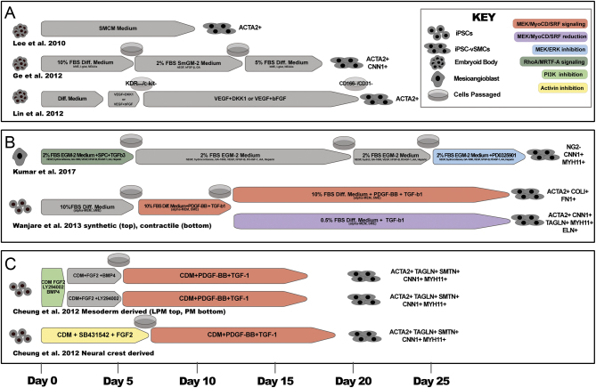
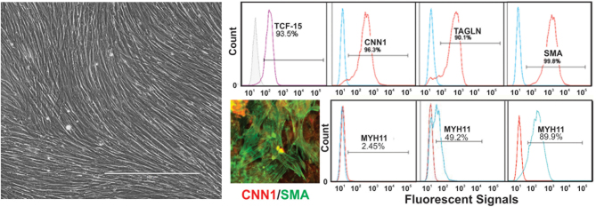
The reproducible generation of human-induced pluripotent stem cell (hiPSC)-derived vascular smooth muscle cells (vSMCs) in vitro has been critical to overcoming many limitations of animal and primary cell models of vascular biology and disease. Since this initial advance, research in the field has turned toward recapitulating the naturally occurring subtype specificity found in vSMCs throughout the body, and honing functional models of vascular disease. In this review, we summarize vSMC derivation approaches, including current phenotype and developmental origin-specific methods, and applications of vSMCs in functional disease models and engineered tissues. Further, we discuss the challenges of heterogeneity in hiPSC-derived tissues and propose approaches to identify and isolate vSMC subtype populations.

Keywords: differentiation; engineered tissues; human iPSC; phenotype switching; vascular smooth muscle cells.


Conflict of interest statement

The authors declare that there is no conflict of interest that could be perceived as prejudicing the impartiality of this review.

Figures

Figure 1
Figure 1
Scaled, graphical summary of selected iPSC-vSMC differentiation protocols. (A) Non-specific differentiation protocols are initiated after formation of an embryoid body (EB) and feature limited vSMC marker characterization. EB differentiation is generally non-directed, but specific cell types can be induced depending on the addition of growth factors at specific times of cultivation (16, 17, 18). (B) Phenotype-driven protocols seek to generate either synthetic or contractile vSMCs through phenotype switching. Cell assessment is made in part through monitoring the formation of contractile structures within cells and levels of MYH11 (30, 37). (C) Lineage-specific differentiation is achieved by way of mesodermal or neuroectoderm intermediates and results in neural crest (NC), lateral plate mesoderm (LPM) or paraxial mesoderm (PM) cell populations (34). Selected pathway targets have been highlighted. The time of cultivation for all of the listed protocols is indicated longitudinally at the bottom of (C) in days. SMCM. smooth muscle cell medium; EGM-2, endothelial growth medium 2; SmGM-2, smooth muscle cell growth medium 2; CDM, chemically defined medium; BME, β-mercaptoethanol; MEM, minimal essential medium; NEAA, non-essential amino acids; EGF, epidermal growth factor; VEGF, vascular endothelial growth factor; FGF2/bFGF, fibroblast growth factor 2; SPC, sphingosylphosphorylcholine; DKK1, Dickkopf 1; PDGF, platelet-derived growth factor; TGF-β1, transforming growth factor beta 1; FBS, fetal bovine serum; ACTA2, α-smooth muscle actin; CNN1, calponin 1; MYH11, smooth muscle myosin heavy chain; NG2, neuron-glial antigen 2; COLI, collagen I; FN1, fibronectin 1; TAGLN, transgelin; ELN, elastin; GA-1000, Gentamicin sulfate-Amphotericin; CD31/PECAM, platelet endothelial cell adhesion molecule; CD166/ALCAM, CD166 antigen or activated leukocyte cell adhesion molecule.
Figure 2
Figure 2
Differentiation of human iPSC line i057 to vSMCs generated via paraxial mesodermal (PM) intermediates. vSMCs derived from iPSCs through PM intermediates are shown here as a monolayer culture cultivated in 2% fetal bovine serum (FBS) (left) (35, 52). The presence of TCF-15-labeled intermediates at differentiation day 7, as well as markers (CNN1, TAGLN and SMA/ACTA2) of differentiated vSMC could be quantified by flow cytometry (top row). Examples showing cell-to-cell and batch-to-batch heterogeneity of vSMCs are shown using co-immunostaining of i057-vSMCs with antibodies to CNN1 and SMA (ACTA2), and by flow cytometry of MYH11 immunostained vSMCs from three independent experiments (right, bottom row).

Similar articles

Cited by

References

    1. Bergers G, Song S. The role of pericytes in blood-vessel formation and maintenance. Neuro-Oncology 2005. 452–464. (10.1215/S1152851705000232) - DOI - PMC - PubMed
    1. Majesky MW. Developmental basis of vascular smooth muscle diversity. Arteriosclerosis, Thrombosis, and Vascular Biology 2007. 1248–1258. (10.1161/ATVBAHA.107.141069) - DOI - PubMed
    1. Majesky MW, Dong XR, Regan JN, Hoglund VJ. Vascular smooth muscle progenitor cells: building and repairing blood vessels. Circulation Research 2011. 365–377. (10.1161/CIRCRESAHA.110.223800) - DOI - PMC - PubMed
    1. Brozovich FV, Nicholson CJ, Degen CV, Gao YZ, Aggarwal M, Morgan KG. Mechanisms of vascular smooth muscle contraction and the basis for pharmacologic treatment of smooth muscle disorders. Pharmacological Reviews 2016. 476–532. (10.1124/pr.115.010652) - DOI - PMC - PubMed
    1. Lim S, Park S. Role of vascular smooth muscle cell in the inflammation of atherosclerosis. BMB Reports 2014. 1–7. (10.5483/bmbrep.2014.47.1.285) - DOI - PMC - PubMed

LinkOut - more resources