Review
. 2020 Feb;2(2):123-136.
doi: 10.1016/j.trechm.2019.07.002.

The Structure and Function of DNA G-Quadruplexes

Affiliations
Review

The Structure and Function of DNA G-Quadruplexes

Jochen Spiegel et al. Trends Chem. 2020 Feb.

Abstract

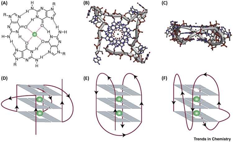
Guanine-rich DNA sequences can fold into four-stranded, noncanonical secondary structures called G-quadruplexes (G4s). G4s were initially considered a structural curiosity, but recent evidence suggests their involvement in key genome functions such as transcription, replication, genome stability, and epigenetic regulation, together with numerous connections to cancer biology. Collectively, these advances have stimulated research probing G4 mechanisms and consequent opportunities for therapeutic intervention. Here, we provide a perspective on the structure and function of G4s with an emphasis on key molecules and methodological advances that enable the study of G4 structures in human cells. We also critically examine recent mechanistic insights into G4 biology and protein interaction partners and highlight opportunities for drug discovery.

Keywords: DNA; G-quadruplex; G4; drug discovery; nucleic acids; secondary structure.


Figures

Figure 1
Figure 1
G-Quadruplex (G4) Structures. (A) Structure of a G-quartet formed by the Hoogsteen hydrogen-bonded guanines and central cation (coloured green) coordinated to oxygen atoms. Crystal structure of human telomeric G4s (Protein Data Bank: 1KF1): (B) top view and (C) side view; backbone is represented by grey tube and the structures are colour-coded by atoms. Schematic representation of unimolecular G4s based on the strand direction: (D) parallel, (E) anti-parallel, and (F) hybrid with a bulge.
Figure 2
Figure 2
G-Quadruplex (G4) Ligands. (A) Crystal structure of a naphthalene diimide, MM41 bound to an intramolecular human telomeric DNA G4, colour-coded by atoms; water molecules are shown as red spheres, MM41 carbon atoms are coloured green, surface of the G4 is coloured light grey (Protein Data Bank: 3UYH). (B) Structures of selected widely used G4 ligands.
Figure 3
Figure 3
Detection and Mapping of DNA G-Quadruplexes (G4s). (A) Visualisation of G4s in fixed or live cells using structure-specific antibodies as well as labelled or intrinsically fluorescent G4 ligands. The number of detected G4 foci can be increased by small molecule treatment or helicase depletion. (B) High-throughput sequencing of G4s in human genomic DNA (G4-Seq). Two consecutive sequencing runs, under normal and G4 stabilising conditions, provide a reference map and detect G4-dependent polymerase stalling, respectively. (C) Chromatin immunoprecipitation employing antibodies against endogenous G4-binding proteins followed by next-generation sequencing (ChIP-seq). Genomic occupancy of endogenous proteins is used to infer putative G4 sites. (D) Treatment with G4 ligands induces G4-dependent DNA damage. ChIP-seq of DNA damage markers in combination with deep sequencing detects G4-associated regions. (E) Permanganate oxidation of nucleotides in transiently unwound regions traps the unpaired state, resulting in sensitivity to a single-strand specific nuclease. Computational prediction is then used to infer the type of underlying non-B-DNA structures based on sequence context. (F) G4-specific chromatin immunoprecipitation and next-generation sequencing (G4 ChIP-seq). A G4-specific antibody (e.g., BG4) is used to precipitate G4 structures directly from native chromatin and is identified by deep sequencing.
Figure 4
Figure 4
Crystal Structure of DHX36 Bound to the c-Myc G4 (Protein Data Bank: 5VHE). Overall structure is shown in cartoon representation with the domain organisation of DHX36 colour-coded (top). The α-helical DHX36-specific motif (DSM) stacks on the 5′-quartet. The residues Ile65, Tyr69, and Ala70 form a nonpolar surface similar to the proposed binding mode of most small molecule G-quadruplex (G4) ligands (bottom).
Figure 5
Figure 5
G-Quadruplex (G4) Can Induce Epigenetic Reprogramming. Unresolved G4 structures (e.g., G4 ligand treatment, impaired helicases) on the leading strand may promote uncoupling of DNA synthesis from opening of the replication fork and disrupt histone recycling. Original histone modifications (blue) are lost and replaced with new histones (brown), resulting in epigenetic reprogramming upstream of G4 sites.

References

    1. Watson J.D., Crick F.H. Molecular structure of nucleic acids; a structure for deoxyribose nucleic acid. Nature. 1953;171:737–738. - PubMed
    1. Bang I. Untersuchungen über die Guanylsäure. Biochem. Z. 1910;26:293–311. (in German)
    1. Gellert M. Helix formation by guanylic acid. Proc. Natl. Acad. Sci. U. S. A. 1962;48:2013–2018. - PMC - PubMed
    1. Arnott S. Structures for polyinosinic acid and polyguanylic acid. Biochem. J. 1974;141:537–543. - PMC - PubMed
    1. Zimmerman S.B. X-ray fiber diffraction and model-building study of polyguanylic acid and polyinosinic acid. J. Mol. Biol. 1975;92:181–192. - PubMed

LinkOut - more resources