

Type I collagen hydrogels as a delivery matrix for royal jelly derived extracellular vesicles
- PMID: 32924637
- PMCID: PMC7534280
- DOI: 10.1080/10717544.2020.1818880
Type I collagen hydrogels as a delivery matrix for royal jelly derived extracellular vesicles
Abstract
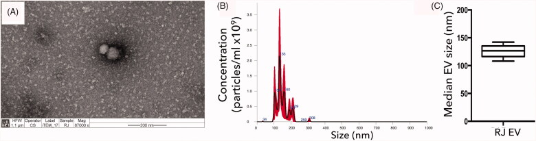
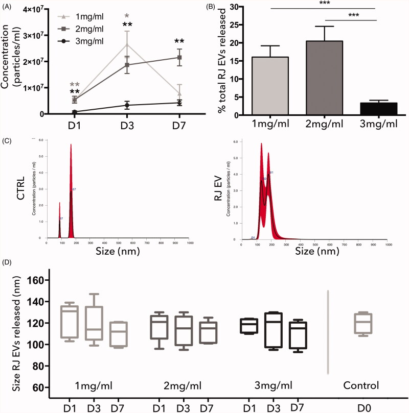
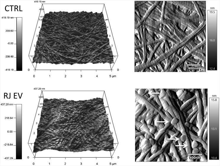
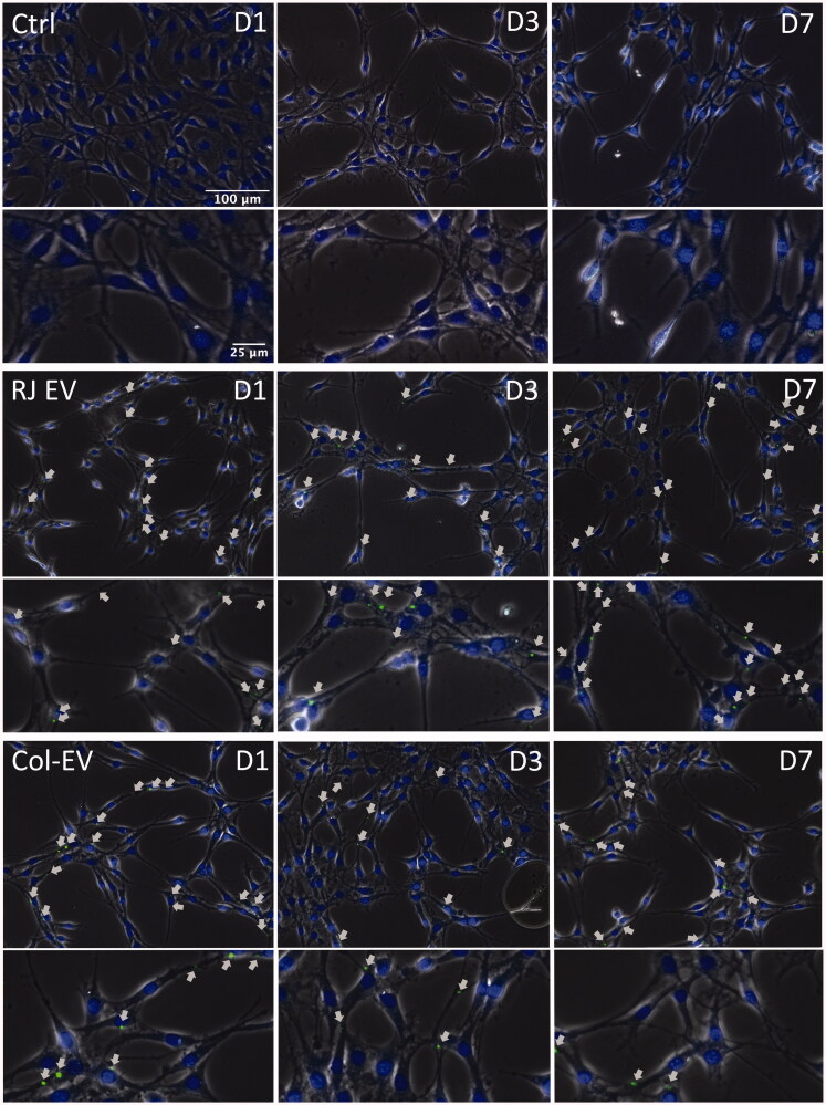
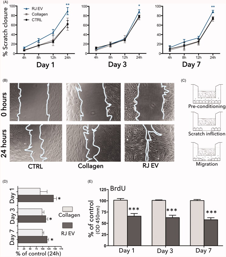
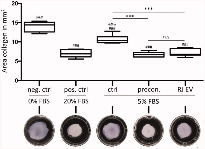
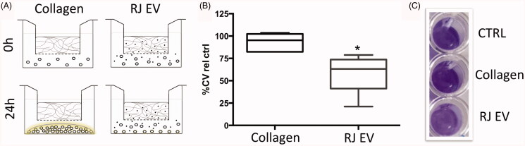
Throughout the last decade, extracellular vesicles (EVs) have become increasingly popular in several areas of regenerative medicine. Recently, Apis mellifera royal jelly EVs (RJ EVs) were shown to display favorable wound healing properties such as stimulation of mesenchymal stem cell migration and inhibition of staphylococcal biofilms. However, the sustained and effective local delivery of EVs in non-systemic approaches - such as patches for chronic cutaneous wounds - remains an important challenge for the development of novel EV-based wound healing therapies. Therefore, the present study aimed to assess the suitability of type I collagen -a well-established biomaterial for wound healing - as a continuous delivery matrix. RJ EVs were integrated into collagen gels at different concentrations, where gels containing 2 mg/ml collagen were found to display the most stable release kinetics. Functionality of released RJ EVs was confirmed by assessing fibroblast EV uptake and migration in a wound healing assay. We could demonstrate reliable EV uptake into fibroblasts with a sustained pro-migratory effect for up to 7 d. Integrating fibroblasts into the RJ EV-containing collagen gel increased the contractile capacity of these cells, confirming availability of RJ EVs to fibroblasts within the collagen gel. Furthermore, EVs released from collagen gels were found to inhibit Staphylococcus aureus ATCC 29213 biofilm formation. Overall, our results suggest that type I collagen could be utilized as a reliable, reproducible release system to deliver functional RJ EVs for wound healing therapies.
Keywords: Apis mellifera; Wound healing; drug delivery; extracellular vesicle delivery; regenerative medicine.
Conflict of interest statement
The authors report no conflicts of interest. The authors alone are responsible for the content and writing of this article.
Figures
Similar articles
-
Multifunctional hydrogel-based engineered extracellular vesicles delivery for complicated wound healing.Theranostics. 2024 Jul 8;14(11):4198-4217. doi: 10.7150/thno.97317. eCollection 2024. Theranostics. 2024. PMID: 39113809 Free PMC article. Review.
-
Extracellular Vesicle-Based Hydrogels for Wound Healing Applications.Int J Mol Sci. 2023 Feb 18;24(4):4104. doi: 10.3390/ijms24044104. Int J Mol Sci. 2023. PMID: 36835516 Free PMC article. Review.
-
In situ formed scaffold with royal jelly-derived extracellular vesicles for wound healing.Theranostics. 2023 May 8;13(9):2811-2824. doi: 10.7150/thno.84665. eCollection 2023. Theranostics. 2023. PMID: 37284440 Free PMC article.
-
Controlled delivery of HIF-1α via extracellular vesicles with collagen-binding activity for enhanced wound healing.J Control Release. 2025 Apr 10;380:330-347. doi: 10.1016/j.jconrel.2025.02.010. Epub 2025 Feb 8. J Control Release. 2025. PMID: 39921033
-
Royal jelly extracellular vesicles promote wound healing by modulating underlying cellular responses.Mol Ther Nucleic Acids. 2023 Feb 14;31:541-552. doi: 10.1016/j.omtn.2023.02.008. eCollection 2023 Mar 14. Mol Ther Nucleic Acids. 2023. PMID: 36895953 Free PMC article.
Cited by
-
An ECM-Mimetic Hydrogel to Promote the Therapeutic Efficacy of Osteoblast-Derived Extracellular Vesicles for Bone Regeneration.Front Bioeng Biotechnol. 2022 Mar 30;10:829969. doi: 10.3389/fbioe.2022.829969. eCollection 2022. Front Bioeng Biotechnol. 2022. PMID: 35433655 Free PMC article.
-
Application of Collagen-Based Hydrogel in Skin Wound Healing.Gels. 2023 Feb 27;9(3):185. doi: 10.3390/gels9030185. Gels. 2023. PMID: 36975634 Free PMC article. Review.
-
Multifunctional hydrogel-based engineered extracellular vesicles delivery for complicated wound healing.Theranostics. 2024 Jul 8;14(11):4198-4217. doi: 10.7150/thno.97317. eCollection 2024. Theranostics. 2024. PMID: 39113809 Free PMC article. Review.
-
Collagen binding properties separate two functionally distinct subpopulations of milk extracellular vesicles regarding bone regenerative capacity.Mater Today Bio. 2025 Jul 18;33:102115. doi: 10.1016/j.mtbio.2025.102115. eCollection 2025 Aug. Mater Today Bio. 2025. PMID: 40727082 Free PMC article.
-
Extracellular Vesicle-Based Hydrogels for Wound Healing Applications.Int J Mol Sci. 2023 Feb 18;24(4):4104. doi: 10.3390/ijms24044104. Int J Mol Sci. 2023. PMID: 36835516 Free PMC article. Review.
References
-
- Achilli M, Mantovani D. (2010). Tailoring mechanical properties of collagen-based scaffolds for vascular tissue engineering: the effects of pH, temperature and ionic strength on gelation. Polymers 2:664–80.
-
- Aguayo S, Strange A, Gadegaard N, et al. (2016). Influence of biomaterial nanotopography on the adhesive and elastic properties of Staphylococcus aureus cells. RSC Adv 6:89347–55.
-
- Ban JJ, Lee M, Im W, Kim M. (2015). Low pH increases the yield of exosome isolation. Biochem Biophys Res Commun 461:76–9. - PubMed
-
- Bang C, Thum T. (2012). Exosomes: new players in cell-cell communication. Int J Biochem Cell Biol 44:2060–4. - PubMed
MeSH terms
Substances
LinkOut - more resources
Full Text Sources
Other Literature Sources
Molecular Biology Databases