

A rare case of giant retroperitoneal neurilemmoma
- PMID: 32924705
- PMCID: PMC7493258
- DOI: 10.1177/0300060520935302
A rare case of giant retroperitoneal neurilemmoma
Abstract
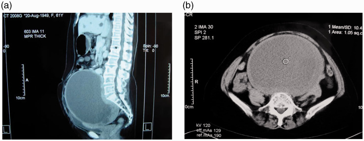
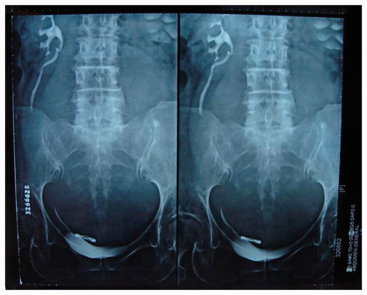
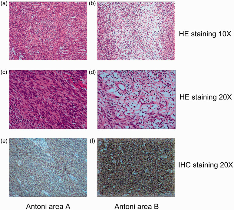
Neurilemmoma, also known as schwannoma or neurinoma, is a tumor that originates from neural sheath Schwann cells. Giant neurilemmomas derived from the retroperitoneum have rarely been reported. We herein describe a woman with a giant retroperitoneal neurilemmoma that was initially incorrectly diagnosed as an inflammatory abdominal mass. The tumor extended from the patient's hypogastrium to her pelvic cavity and measured 20 × 15 × 10 cm. The tumor was excised via laparotomy and diagnosed as a retroperitoneal neurilemmoma through histological and immunohistochemical examination. Although rare, particularly in the giant form, neurilemmoma should be considered as an important differential diagnosis in patients with a retroperitoneal tumor or inflammatory abdominal mass. Complete excision should be considered for the potential cure of giant retroperitoneal neurilemmomas.
Keywords: Giant neurilemmoma; Schwann cells; histology; immunohistochemistry; laparotomy; retroperitoneal tumor; treatment.
Figures
References
-
- Sato Y, Imanishi Y, Tomita T, et al. Clinical diagnosis and treatment outcomes for parapharyngeal space schwannomas: a single-institution review of 21 cases. Head Neck 2018; 40: 569–576. - PubMed
-
- Li S, Lu X, Xie S, et al. Intraparotid facial nerve schwannoma: a 17-year, single-institution experience of diagnosis and management. Acta Otolaryngol 2019; 139: 444–450. - PubMed
-
- Fini G, Leonardi A, Mici E, et al. Schwannoma of the parotid gland. Case report. Ann Ital Chir 2015; 86: 1–4. - PubMed
-
- Hori T, Yamagiwa K, Yagi S, et al. Noradrenalin-secreting retroperitoneal schwannoma resected by hand-assisted laparoscopic surgery: report of a case. Surg Today 2006; 36: 1108–1113. - PubMed
Publication types
MeSH terms
LinkOut - more resources
Full Text Sources
