

MRI predictors for brain invasion in meningiomas
- PMID: 32924772
- PMCID: PMC7868592
- DOI: 10.1177/1971400920953417
MRI predictors for brain invasion in meningiomas
Abstract
Background and purpose: In the 2016 revision of the World Health Organization classification of central nervous system tumours, brain invasion was added as an independent histological criterion for the diagnosis of a World Health Organization grade II atypical meningioma. The aim of this study was to assess whether magnetic resonance imaging characteristics can predict brain invasion for meningiomas.
Materials and methods: We conducted a retrospective review of all meningiomas resected at our institution between 2005 and 2016 which had preoperative magnetic resonance imaging and included brain tissue within the pathology specimen. One hundred meningiomas were included in the study, 60 of which had histopathological brain invasion, 40 of which did not. Magnetic resonance imaging characteristics of tumours were evaluated for potential predictors of brain invasion. Tumour location, size, perilesional oedema, contour, cerebrospinal fluid cleft, peritumoral cyst, dural venous sinus invasion, bone invasion, hyperostosis and the presence of enlarged pial arteries and veins were evaluated. Data were analysed using conventional chi-square, Fisher's exact test and logistic regression.
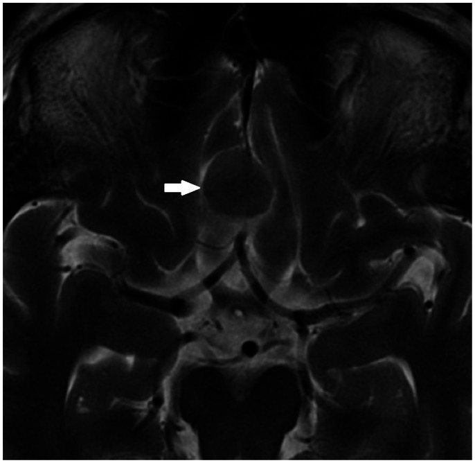
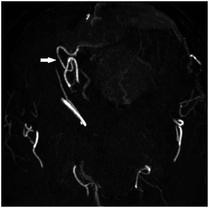
Results: The volume of peritumoral oedema was significantly higher in the brain-invasive meningioma group compared to the non-brain-invasive group. The presence of a complete cleft was a rare finding that was only found in non-brain-invasive meningiomas. The presence of enlarged pial feeding arteries was a rare finding that was only found in brain-invasive meningiomas.
Conclusions: An increased volume of perilesional oedema is associated with the likelihood of brain invasion for meningiomas.
Keywords: MRI; Meningioma; brain invasion.
Figures
References
-
- Chernov MF, Kasuya H, Nakaya K, et al. 1H-MRS of intracranial meningiomas: what it can add to known clinical and MRI predictors of the histopathological and biological characteristics of the tumor? Clin Neurol Neurosurg 2011; 113: 202–212. - PubMed
-
- Johnson DR, Guerin JB, Giannini C, et al. 2016 Updates to the WHO Brain Tumor Classification System: what the radiologist needs to know. RadioGraphics 2017; 37: 2164--2180. - PubMed
MeSH terms
LinkOut - more resources
Full Text Sources
Medical
