. 2022 Feb;40(2):903-917.
doi: 10.1080/07391102.2020.1819881. Epub 2020 Sep 14.

Potential protease inhibitors and their combinations to block SARS-CoV-2

Affiliations

Potential protease inhibitors and their combinations to block SARS-CoV-2

Chandran S Abhinand et al. J Biomol Struct Dyn. 2022 Feb.

Abstract

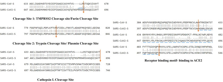
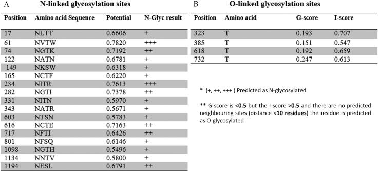
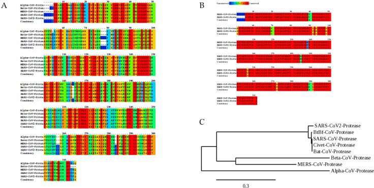
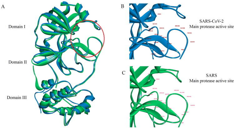
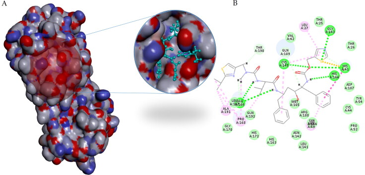
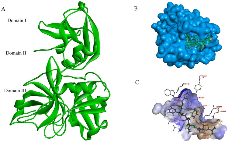
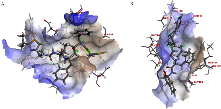
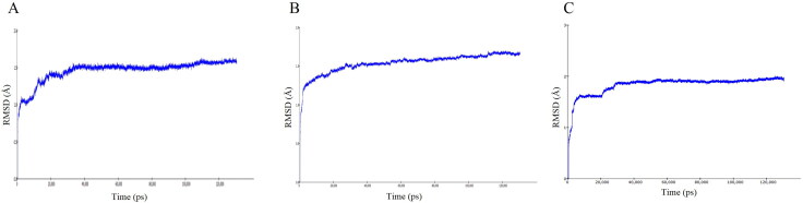
COVID-19, which has emerged recently as a pandemic viral infection caused by SARS-coronavirus 2 has spread rapidly around the world, creating a public health emergency. The current situation demands an effective therapeutic strategy to control the disease using drugs that are approved, or by inventing new ones. The present study examines the possible repurposing of existing anti-viral protease inhibitor drugs. For this, the structural features of the viral spike protein, the substrate for host cell protease and main protease of the available SARS CoV-2 isolates were established by comparing with related viruses for which antiviral drugs are effective. The results showed 97% sequence similarity among SARS and SARS-CoV-2 main protease and has same cleavage site positions and ACE2 receptor binding region as in the SARS-CoV spike protein. Though both are N-glycosylated, unlike SARS-CoV, human SARS-CoV-2 S-protein was O-glycosylated as well. Molecular docking studies were done to explore the role of FDA approved protease inhibitors to control SARS-CoV-2 replication. The results indicated that, Ritonavir has the highest potency to block SARS-CoV-2 main protease and human TMPRSS2, a host cell factor that aids viral infection. Other drugs such as Indinavir and Atazanavir also showed favourable binding with Cathepsin B/L that helped viral fusion with the host cell membrane. Further molecular dynamics simulation and MM-PBSA binding free energy calculations confirmed the stability of protein-drug complexes. These results suggest that protease inhibitors particularly Ritonavir, either alone or in combination with other drugs such as Atazanavir, have the potential to treat COVID 19.Communicated by Ramaswamy H. Sarma.

Keywords: COVID-19; TMPRSS2; main protease; molecular docking; spike protein.


Conflict of interest statement

The authors declare no conflict of interest.

Figures

Figure 1.
Figure 1.
Schematic representation of SARS-CoV-2 spike protein with cleavage sites and receptor binding sites. The S protein sequence of SARS-CoV and SARS-CoV-2 cleavage sites were aligned using EMBOSS Needle tool. Cleavage site 1(TMPRSS2/Furin), Cleavage site 2(Trypsin/Plasmin) and ACE2 binding sites corresponding to SARS-CoV and SARS-CoV-2 spike proteins are indicated by coloured lines. Polybasic furin recognition site ‘RRAR’ in S1 cleavage site is highlighted with blue colour lines.
Figure 2.
Figure 2.
Glycosylation sites of SARS-CoV-2 spike protein (A) N-glycosylation sites of SARS-Cov-2 spike protein (NCBI Reference Sequence: YP_009724390.1) was predicted using NetNGlyc 1.0 Server. 17 N-linked glycosylation sites were predicted with threshold value > 0.5. (B) O-linked glycosylation sites of SARS-CoV-2 spike protein (NCBI Reference Sequence: YP_009724390.1) was predicted using NetOGlyc 3.1 server and the results showed 4 threonine glycosylation sites with G-score < 0.5, I-score > 0.5 and no predicted neighbouring residues with in a distance ten.
Figure 3.
Figure 3.
Phylogenetic and sequence alignment of SARS-CoV2protease. (A) Multiple sequence alignment showing conserved and non-conserved regions in main protease of different corona virus strains [(Alpha coronaviruses -NL63: PDB ID-3TLO), (Beta coronavirus- HKU1: PDB ID-3D23), (SARS-CoV: PDB ID-2AMQ), (MERS-CoV: PDB ID-4WMD), (SARS-CoV-2:PDB ID-6LU7)]. Sequence alignment was done using PRALINE tool and observed some active site residues similarities among all the strains. Blue to red color indicates un-conserved residues to conserved residues. (B) Sequence alignment between SARS and SARS-CoV-2 proteases. Pairwise sequence alignment revealed more than 97% sequence similarities among the sequences. (C) Phylogenetic tree of SARS related main protease. Phylogenetic tree was constructed using phelogeny.fr. and identified that bat- derived SARS main protease is conspecific with human SARS-CoV-2 main protease.
Figure 4.
Figure 4.
Comparison of crystal structures of main protease from SARS-CoV (PDB ID-2AMQ) and SARS-CoV-2 (PDB ID-6LU7). (A) Superimposition of SARS (green colour) and SARS-CoV-2 (blue colour) main proteases using 2020. Domains are labeled by numbers and the residues 8-101, 102-184, 201-303 represent domain I, II and III respectively. (B) Active site residues labelled in SARS-CoV-2 main protease. (C) Active site residues labelled in SARS main protease.
Figure 5.
Figure 5.
Docking of SARS-CoV-2 main protease against Ritonavir. Molecular docking of SARS-CoV-2 main protease (PDB ID-6LU7) against Ritonavir (CID_392622) showed highest docking score and favourable intermolecular interactions with in the 16 ligands. The image is a representative of docked pose with highest docking score (A) Ritonavir binds in the active site of SARS-CoV-2 main protease. (B) 2 D interaction map of H-bonds formed between Ritonavir and amino acids in SARS-CoV-2 protease.
Figure 6.
Figure 6.
Modelled structure of TMPRSS2 and docking of TMPRSS2 protease against Ritonavir. (A) Homology modelled 3D structure of TMPRSS2 using Discovery studio 2020. Domains are labeled by numbers and the residues 1-84, 85-105, 106-492 represent domain I, II and III respectively. Amino acid residues His296, Asp345, Ser441 and Asp435 which constitute the active site were located in domain III (B) Ritonavir (CID_392622) binds in the active site of TMPRSS2 protease. The image is a representative of docked pose with highest docking score (C) Intermolecular interactions formed between Ritonavir and amino acids in TMPRSS2 protease.
Figure 7.
Figure 7.
Docking view of Atazanavir in binding site of Cathepsin L (A) and Indinavir in binding site of Cathepsin B (B). Intermolecular interactions between the receptor in surface view and ligand corresponding to each interactions were drawn in Discovery studio 2020. Molecular docking of Cathepsin L (PDB ID- 2XU1) against Atazanavir (CID_148192) and Cathepsin B (PDB ID- 1CSB) against Indinavir (CID_5362440) showed highest docking score and favourable intermolecular interactions with in the 16 ligands. Each image is a representative of docked pose of the respective ligand with highest docking score.
Figure 8.
Figure 8.
RMSD plot of top ranked protease inhibitors complexed with three different targets. (A) RMSD of SARS-CoV-2 main protease- Ritonavir complex showed that the structure was stable around 1.50 Å for about 130 ns. (B) RMSD of TMPRSS2- Ritonavir complex showed the complex was stable around 1.40 Å for about 130 ns. (C) Cathepsin L- Atazanavir complex showed the complex was stable around 1.85 Å for about 130 ns.
Figure 9.
Figure 9.
RMSF plot of three different targets with potential protease inhibitors. (A) RMSF plot of SARS-CoV-2 main protease- Ritonavir complex showed the amino acid residue fluctuation is within 1.3 Å. (B) RMSF plot of TMPRSS2- Ritonavir complex showed the residue fluctuation is within 1.7 Å.(C) RMSF plot of Cathepsin L- Atazanavir complex showed the fluctuation is within 1.3 Å.
Figure 10.
Figure 10.
MM-PBSA of protein -ligand complexes at different time intervals. MM_PBSA calculations were done by binding energy calculation protocol in Discovery Studio 2020. A-Main protease –Ritonavir complex B-TMPRSS2-Ritonavir complex, C-Cathepsin-L-Atazanavir complex. Results given are the mean of multiple binding free energy values (in units of kcal/mol) ±SEM at 10 ns intervals. Comparison of the mean values of each complex at different time intervals was made by one way ANOVA analysis. p > 0.05 not significant.
Figure 11.
Figure 11.
Heat map representing the consistency among the hydrogen bonds formed in three complexes. (A) SARS-CoV-2 main protease- Ritonavir complex showed four consistent hydrogen bonds during the simulation. (B) TMPRSS2- Ritonavir complex showed two consistent hydrogen bonds during the simulation. (C) Cathepsin L- Atazanavir complex showed six consistent hydrogen bonds during the simulation.

References

    1. Bagdonaite, I., Nordén, R., Joshi, H. J., Dabelsteen, S., Nyström, K., Vakhrushev, S. Y., Olofsson, S., & Wandall, H. H. (2015). A strategy for O-glycoproteomics of enveloped viruses-the O-glycoproteome of herpes simplex virus type 1. PLoS Pathogens, 11(4), e1004784. 10.1371/journal.ppat.1004784 - DOI - PMC - PubMed
    1. Bhardwaj, V. K., Singh, R., Sharma, J., Rajendran, V., Purohit, R., & Kumar, S. (2020). Identification of bioactive molecules from tea plant as SARS-CoV-2 main protease inhibitors. Journal of Biomolecular Structure and Dynamics,1–10. 10.1080/07391102.2020.1766572 - DOI - PMC - PubMed
    1. Cao, B., Wang, Y., Wen, D., Liu, W., Wang, J., Fan, G., Ruan, L., Song, B., Cai, Y., Wei, M., Li, X., Xia, J., Chen, N., Xiang, J., Yu, T., Bai, T., Xie, X., Zhang, L., Li, C., … Wang, C. (2020). A trial of lopinavir-ritonavir in adults hospitalized with severe covid-19. The New England Journal of Medicine, 382(19), 1787–1799. 10.1056/NEJMoa2001282 - DOI - PMC - PubMed
    1. Chan, K. S., Lai, S. T., Chu, C. M., Tsui, E., Tam, C. Y., & Wong, M. M. (2003). Treatment of severe acute respiratory syndrome with lopinavir/ritonavir: A multicentre retrospective matched cohort study. Hong Kong Medical Journal, 9(6), 399–406. - PubMed
    1. Chen, J., Lee, K. H., Steinhauer, D. A., Stevens, D. J., Skehel, J. J., & Wiley, D. C. (1998). Structure of the hemagglutinin precursor cleavage site, a determinant of influenza pathogenicity and the origin of the labile conformation. Cell, 95(3), 409–417. 10.1016/S0092-8674(00)81771-7 - DOI - PubMed

Publication types

Substances