

Reduced Monocytic Human Leukocyte Antigen-DR Expression Indicates Immunosuppression in Critically Ill COVID-19 Patients
- PMID: 32925314
- PMCID: PMC7288784
- DOI: 10.1213/ANE.0000000000005044
Reduced Monocytic Human Leukocyte Antigen-DR Expression Indicates Immunosuppression in Critically Ill COVID-19 Patients
Abstract
Background: The cellular immune system is of pivotal importance with regard to the response to severe infections. Monocytes/macrophages are considered key immune cells in infections and downregulation of the surface expression of monocytic human leukocyte antigen-DR (mHLA-DR) within the major histocompatibility complex class II reflects a state of immunosuppression, also referred to as injury-associated immunosuppression. As the role of immunosuppression in coronavirus disease 2019 (COVID-19) is currently unclear, we seek to explore the level of mHLA-DR expression in COVID-19 patients.
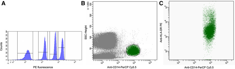
Methods: In a preliminary prospective monocentric observational study, 16 COVID-19-positive patients (75% male, median age: 68 [interquartile range 59-75]) requiring hospitalization were included. The median Acute Physiology and Chronic Health Evaluation-II (APACHE-II) score in 9 intensive care unit (ICU) patients with acute respiratory failure was 30 (interquartile range 25-32). Standardized quantitative assessment of HLA-DR on monocytes (cluster of differentiation 14+ cells) was performed using calibrated flow cytometry at baseline (ICU/hospital admission) and at days 3 and 5 after ICU admission. Baseline data were compared to hospitalized noncritically ill COVID-19 patients.
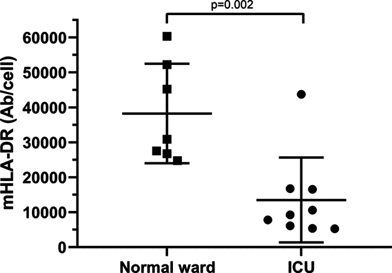
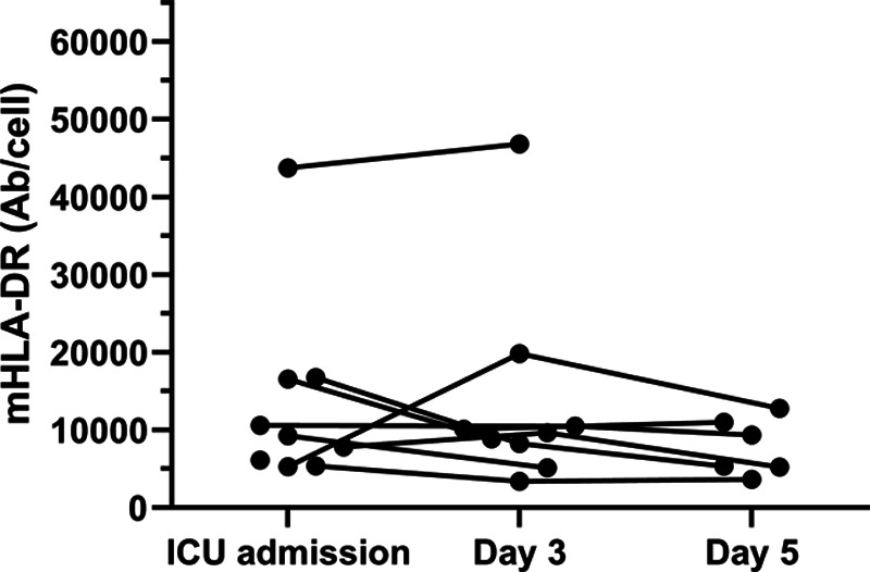
Results: While normal mHLA-DR expression was observed in all hospitalized noncritically ill patients (n = 7), 89% (8 of 9) critically ill patients with COVID-19-induced acute respiratory failure showed signs of downregulation of mHLA-DR at ICU admission. mHLA-DR expression at admission was significantly lower in critically ill patients (median, [quartiles]: 9280 antibodies/cell [6114, 16,567]) as compared to the noncritically ill patients (30,900 antibodies/cell [26,777, 52,251]), with a median difference of 21,508 antibodies/cell (95% confidence interval [CI], 14,118-42,971), P = .002. Reduced mHLA-DR expression was observed to persist until day 5 after ICU admission.
Conclusions: When compared to noncritically ill hospitalized COVID-19 patients, ICU patients with severe COVID-19 disease showed reduced mHLA-DR expression on circulating CD14+ monocytes at ICU admission, indicating a dysfunctional immune response. This immunosuppressive (monocytic) phenotype remained unchanged over the ensuing days after ICU admission. Strategies aiming for immunomodulation in this population of critically ill patients should be guided by an immune-monitoring program in an effort to determine who might benefit best from a given immunological intervention.
Conflict of interest statement
Conflicts of Interest: See Disclosures at the end of the article.
Figures
Comment in
-
Human Leukocyte Antigen-DR Deficiency and Immunosuppression-Related End-Organ Failure in SARS-CoV2 Infection.Anesth Analg. 2020 Oct;131(4):989-992. doi: 10.1213/ANE.0000000000005140. Anesth Analg. 2020. PMID: 32925313 Free PMC article. No abstract available.
Similar articles
-
Case Report: Interferon-γ Restores Monocytic Human Leukocyte Antigen Receptor (mHLA-DR) in Severe COVID-19 With Acquired Immunosuppression Syndrome.Front Immunol. 2021 Apr 7;12:645124. doi: 10.3389/fimmu.2021.645124. eCollection 2021. Front Immunol. 2021. PMID: 33897692 Free PMC article.
-
Predicting the Outcomes of Subjects With Severe Community-Acquired Pneumonia Using Monocyte Human Leukocyte Antigen-DR.Respir Care. 2015 Nov;60(11):1635-42. doi: 10.4187/respcare.03953. Epub 2015 Aug 11. Respir Care. 2015. PMID: 26264418
-
Association of monocyte HLA-DR expression over time with secondary infection in critically ill children: a prospective observational study.Eur J Pediatr. 2022 Mar;181(3):1133-1142. doi: 10.1007/s00431-021-04313-7. Epub 2021 Nov 10. Eur J Pediatr. 2022. PMID: 34755207 Free PMC article.
-
Monocyte HLA-DR Measurement by Flow Cytometry in COVID-19 Patients: An Interim Review.Cytometry A. 2020 Dec;97(12):1217-1221. doi: 10.1002/cyto.a.24249. Epub 2020 Nov 4. Cytometry A. 2020. PMID: 33125816 Review.
-
[Low monocytic HLA-DR expression and risk of secondary infection].Ann Fr Anesth Reanim. 2010 May;29(5):368-76. doi: 10.1016/j.annfar.2010.02.015. Epub 2010 Mar 30. Ann Fr Anesth Reanim. 2010. PMID: 20356708 Review. French.
Cited by
-
Flow cytometry-based high-throughput RNAi screening for miRNAs regulating MHC class II HLA-DR surface expression.Eur J Immunol. 2022 Sep;52(9):1452-1463. doi: 10.1002/eji.202149735. Epub 2022 Jun 9. Eur J Immunol. 2022. PMID: 35612261 Free PMC article.
-
SARS-CoV-2 infection: Understanding the immune system abnormalities to get an adequate diagnosis.Bosn J Basic Med Sci. 2021 Oct 1;21(5):503-514. doi: 10.17305/bjbms.2020.5400. Bosn J Basic Med Sci. 2021. PMID: 33596401 Free PMC article. Review.
-
The dynamics of inflammatory markers in coronavirus disease-2019 (COVID-19) patients: A systematic review and meta-analysis.Clin Epidemiol Glob Health. 2021 Jul-Sep;11:100727. doi: 10.1016/j.cegh.2021.100727. Epub 2021 Mar 20. Clin Epidemiol Glob Health. 2021. PMID: 33778183 Free PMC article. Review.
-
Human Leukocyte Antigen-DR Deficiency and Immunosuppression-Related End-Organ Failure in SARS-CoV2 Infection.Anesth Analg. 2020 Oct;131(4):989-992. doi: 10.1213/ANE.0000000000005140. Anesth Analg. 2020. PMID: 32925313 Free PMC article. No abstract available.
-
Longitudinal single cell atlas identifies complex temporal relationship between type I interferon response and COVID-19 severity.Nat Commun. 2024 Jan 18;15(1):567. doi: 10.1038/s41467-023-44524-0. Nat Commun. 2024. PMID: 38238298 Free PMC article.
References
-
- Rieckmann JC, Geiger R, Hornburg D. Social network architecture of human immune cells unveiled by quantitative proteomics. Nat Immunol. 2017;18:583–593. - PubMed
-
- Rubio I, Osuchowski MF, Shankar-Hari M. Current gaps in sepsis immunology: new opportunities for translational research. Lancet Infect Dis. 2019;19:e422–e436. - PubMed
-
- Schefold JC, Hasper D, Reinke P, Monneret G, Volk HD. Consider delayed immunosuppression into the concept of sepsis. Crit Care Med. 2008;36:3118. - PubMed
MeSH terms
Substances
LinkOut - more resources
Full Text Sources
Other Literature Sources
Research Materials
