. 2020 Sep 1;61(11):19.
doi: 10.1167/iovs.61.11.19.

S100A4 Silencing Facilitates Corneal Wound Healing After Alkali Burns by Promoting Autophagy via Blocking the PI3K/Akt/mTOR Signaling Pathway

Affiliations

S100A4 Silencing Facilitates Corneal Wound Healing After Alkali Burns by Promoting Autophagy via Blocking the PI3K/Akt/mTOR Signaling Pathway

Yulin Wang et al. Invest Ophthalmol Vis Sci. .

Erratum in

Abstract

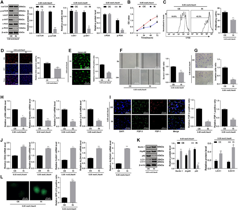
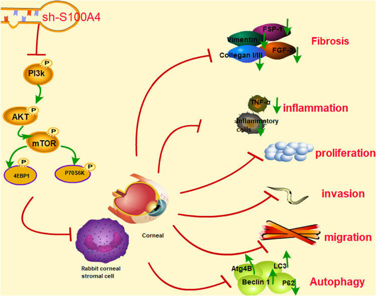
Purpose: This study investigated the role of S100 calcium binding protein A4 (S100A4) in corneal wound healing and the underlying mechanism of the S100A4-mediated PI3K/Akt/mammalian target of rapamycin (mTOR) pathway.

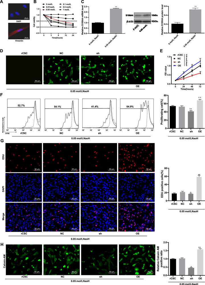
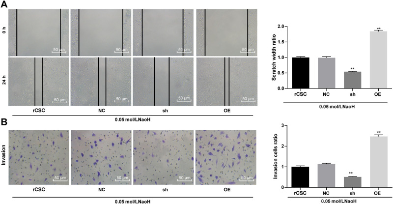
Methods: The rabbit corneal alkali burn model was established in vivo. S100A4 expression, wound healing, inflammation, and autophagy in rabbit cornea after alkali burn were detected. The NaOH-treated rabbit corneal stromal cells (rCSCs) were transfected with overexpressed S100A4 or silencing S100A4 to examine the effect of S100A4 on corneal wound healing in vitro. The effect of S100A4 on cell viability, proliferation, migration, invasion, fibrosis, and autophagy of rCSCs after alkali burn was analyzed. Then the functional rescue experiments were carried out. The PI3K inhibitor, LY294002, was used to elucidate the PI3K/Akt/mTOR signaling pathway in rCSCs.

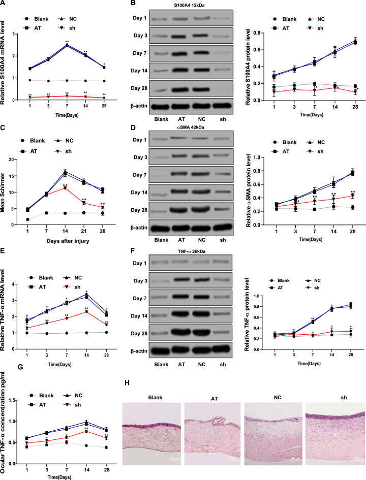
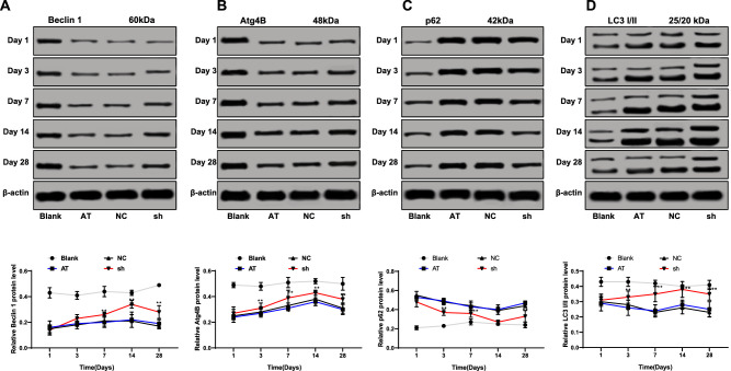
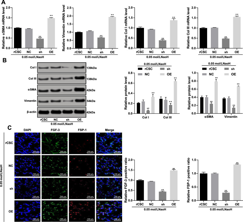
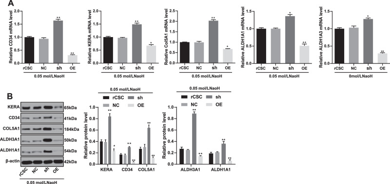
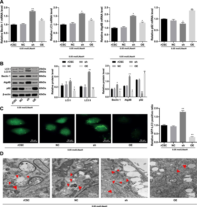
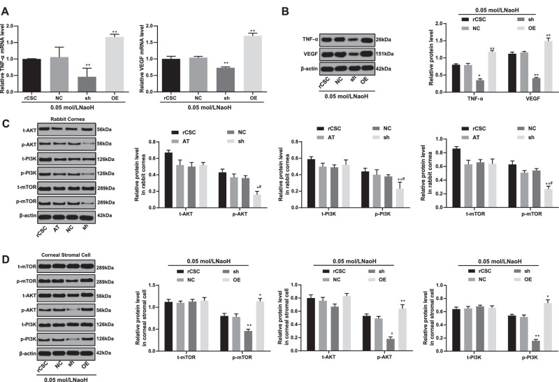
Results: S100A4 silencing promoted rabbit corneal wound healing by inhibiting fibrosis and inflammation and promoting autophagy in alkali-burned cornea, corresponding to increased levels of LC3, Beclin 1, and Atg4B but lowered α-smooth muscle actin, TNF-ɑ, and p62 levels. Moreover, silencing S100A4 inhibited proliferation, migration, invasion, and fibrosis of NaOH-treated rCSCs and promoted the differentiation of rCSCs into corneal cells and the autophagy of damaged rCSCs. The inhibitory role of S100A4 in wound healing was achieved via activation of the PI3K/Akt/mTOR pathway.

Conclusions: S100A4 silencing confers a promising effect on wound healing of alkali-burned cornea by blocking the PI3K/Akt/mTOR pathway, supporting the advancement of corneal gene therapies for wound healing.


Conflict of interest statement

Disclosure: Y. Wang, None; G. Gao, None; Y. Wu, None; Y. Wang, None; X. Wu, None; Q. Zhou, None

Figures

Figure 1.
Figure 1.
Silencing S100A4 contributed to promotion of corneal wound healing after alkali burn. The mRNA (A) and protein (B) levels of S100A4 (S100A4) in rabbit cornea measured by RT-qPCR and Western blot assay; formation of tears detected using the Schirmer test (C); the protein level of ɑ-SMA in rabbit cornea measured by Western blot assay (D); the mRNA (E) and protein (F) levels of TNF-ɑ (TNF-ɑ) in rabbit cornea measured by RT-qPCR and Western blot assay; the protein concentration of TNF-ɑ in aqueous humor measured by ELISA assay (G); hematoxylin and eosin staining was carried out on the cornea to observe the inflammatory cells in cornea at day 28 (H). n = 3. On days 1, 3, 7, 14, and 28, three New Zealand white rabbits from each group were used for the Schirmer test, and corneal tissue was extracted for RT-qPCR and Western blot assay. Data are expressed as mean ± standard deviation. Two-way ANOVA and Tukey's multiple comparisons test were used for data analysis. *P < 0.05. **P < 0.01.
Figure 2.
Figure 2.
Silencing of S100A4 contributed to promotion of autophagy after corneal alkali burn in rabbits. The protein levels of LC3, Beclin 1 (A), Atg4B (B), p62 (C), and LC3 (D) in the cornea after S100A4 silencing were detected by Western blot assay. On days 1, 3, 7, 14, and 28, corneas of three New Zealand white rabbits from each group were used for Western blot assay. Data are expressed as mean ± standard deviation. Replicates = 3. Two-way ANOVA and Tukey's multiple comparisons test were used for data analysis. *P < 0.05. **P < 0.01.
Figure 3.
Figure 3.
Silencing of S100A4 resulted in reduced viability of rCSCs in a corneal alkali burn model. Detection of rCSCs-specific marker vimentin by immunocytochemistry (A); construction of alkali burn rCSC model using different concentrations of NaOH and detection of rCSC viability by MTT assay (B); mRNA and protein levels of S100A4 (S100A4) in NaOH-treated and normal rCSCs by RT-qPCR and Western blot assay (C); detection of S100A4 transfection efficiency by a fluorescence microscope (D); detection of cell viability using MTT assay (E); rCSCs were stained with CFSE and planted into six-well plates. The cells were collected and detected by flow cytometry after 48 hours (F); detection of cell proliferation by EdU labeling assay (G); calcein-AM staining for viable cells (H). Replicates = 3. Data are expressed as mean ± standard deviation. Two-way or one-way ANOVA and Tukey's multiple comparisons test were used for data analysis. *P < 0.05. **P < 0.01.
Figure 4.
Figure 4.
Silencing of S100A4 suppressed the migration and invasion abilities of rCSCs following corneal alkali burn. S100A4 shRNA vector, overexpressed S100A4 vector, and NC vector were constructed and transfected into rCSCs after alkali burn modeling. The migration and invasion abilities of rCSCs after alkali burn were measured by scratch test (A) and Transwell assay (B). Replicates = 3. Data are expressed as mean ± standard deviation. Two-way or one-way ANOVA and Tukey's multiple comparisons test were used for data analysis.*P < 0.05. **P < 0.01.
Figure 5.
Figure 5.
Silencing of S100A4 suppressed the fibrosis of rCSCs induced by corneal alkali burn. S100A4 shRNA vector, overexpressed S100A4 vector, and NC vector were constructed and transfected into rCSCs after alkali burn modeling. RT-qPCR and Western blot assay showing mRNA (A) and protein (B) levels of ɑ-SMA (ɑ-SMA), vimentin (vimentin), and type I/III collagen in rCSCs after alkali burn; immunofluorescence assay showing FGF-3-positive and FSP-1-positive cells in rCSCs after alkali burn (C). Replicates = 3. Data are expressed as mean ± standard deviation. Two-way or one-way ANOVA and Tukey's multiple comparisons test were used for data analysis. *P < 0.05. **P < 0.01.
Figure 6.
Figure 6.
Silencing of S100A4 induced differentiation of rCSCs following corneal alkali burn. S100A4 shRNA vector, overexpressed S100A4 vector, and NC vector were constructed and transfected into rCSCs after alkali burn modeling. RT-qPCR and Western blot assay showing mRNA (A) and protein (B) levels of CD34 (CD34), keratocan (KERA), COL5A1 (COL5A1), ALDH1A1 (ALDH1A1), and ALDH1A1 (ALDH3A1) in rCSCs in the corneal alkali burn model. Replicates = 3. Data are expressed as mean ± standard deviation. Two-way or one-way ANOVA and Tukey's multiple comparisons test were used for data analysis. *P < 0.05. **P < 0.01.
Figure 7.
Figure 7.
Silencing of S100A4 enhanced the autophagy of rCSCs after alkali burn. S100A4 shRNA vector, overexpressed S100A4 vector, and NC vector were constructed and transfected into rCSCs after alkali burn modeling. The mRNA (A) and protein (B) levels of autophagy-related markers in rCSCs after alkali burn determined by RT-qPCR and Western blot assay; immunofluorescence (C) and Transmission Electron Microscopy (TEM) (D) showing LC3 protein distribution and the formation of autophagosomes. Replicates = 3. Data are expressed as mean ± standard deviation. Two-way or one-way ANOVA and Tukey's multiple comparisons test were used for data analysis. *P < 0.05. **P < 0.01. TEM, Transmission Electron Microscopy.
Figure 8.
Figure 8.
Silencing of S100A4 downregulated the PI3K/Akt/mTOR pathway to inhibit inflammation. The mRNA (A) and protein (B) levels of VEGF (VEGF) and TNF-ɑ (TNF-ɑ) in rCSCs after alkali burn determined by RT-qPCR and Western blot assay; the phosphorylation of PI3K, Akt, and mTOR in rabbit cornea (C) and rCSCs (D) after alkali burn measured by Western blot assay. Replicates = 3. Data are expressed as mean ± standard deviation. Two-way or one-way ANOVA and Tukey's multiple comparisons test were used for data analysis. *P < 0.05. **P < 0.01.
Figure 9.
Figure 9.
S100A4 inhibited corneal wound healing after alkali burn via activation of the PI3K/Akt/mTOR signaling pathway. S100A4 shRNA vector, overexpressed S100A4 vector, and NC vector were constructed and transfected into rCSCs after alkali burn modeling. The phosphorylation of PI3K, Akt, and mTOR measured by Western blot assay (A); cell viability detected by MTT assay (B); rCSCs were stained with CFSE, planted into six-well plates, and detected by flow cytometry after 48 hours (C); cell proliferation detected by EdU labeling assay (D); cell viability detected by calcein-AM staining (E); migration (F) and invasion abilities (G) of rCSCs assessed by scratch test and Transwell assay; mRNA levels of vimentin and E-cadherin detected by RT-qPCR (H); FGF-3 and FSP-1 protein expression measured by immunofluorescence (I); mRNA levels of corneal cell-specific markers (J); levels of autophagy-related markers in rCSCs after alkali burn (K); LC3 protein distribution in rCSCs assessed by immunofluorescence. Replicates = 3. Data are expressed as mean ± standard deviation. Two-way or one-way ANOVA and Tukey's multiple comparisons test were used for data analysis. *P < 0.05. **P < 0.01.
Figure 10.
Figure 10.
Silencing of S100A4 downregulated the PI3K/Akt/mTOR pathway, enhanced autophagy, and promoted wound healing of rabbit cornea after alkali burn. After S100A4 silencing, the phosphorylation of the PI3K/Akt/mTOR was decreased significantly. The inhibition of the PI3K/Akt/mTOR pathway promoted the autophagy of rCSCs after alkali burn, led to the differentiation of rCSCs into corneal cells, and inhibited the proliferation, invasion, and migration of rCSCs, thereby promoting the wound healing after alkali burn.

Similar articles

Cited by

References

    1. Ramponi DR. Chemical burns of the eye. Adv Emerg Nurs J. 2017; 39: 193–198. - PubMed
    1. Ljubimov AV, Saghizadeh M.. Progress in corneal wound healing. Prog Retin Eye Res. 2015; 49: 17–45. - PMC - PubMed
    1. Bian F, Pelegrino FS, Henriksson JT, et al. .. Differential effects of dexamethasone and doxycycline on inflammation and MMP production in murine alkali-burned corneas associated with dry eye. Ocul Surf. 2016; 14: 242–254. - PMC - PubMed
    1. Ziaei M, Greene C, Green CR. Wound healing in the eye: therapeutic prospects. Adv Drug Deliv Rev. 2018; 126: 162–176. - PubMed
    1. Mohan RR, Tovey JC, Sharma A, Tandon A. Gene therapy in the cornea: 2005–present. Prog Retin Eye Res. 2012; 31: 43–64. - PMC - PubMed

Publication types

MeSH terms