

Does microenema administration improve the quality of DWI sequences in rectal MRI?
- PMID: 32926212
- PMCID: PMC7946648
- DOI: 10.1007/s00261-020-02718-w
Does microenema administration improve the quality of DWI sequences in rectal MRI?
Abstract
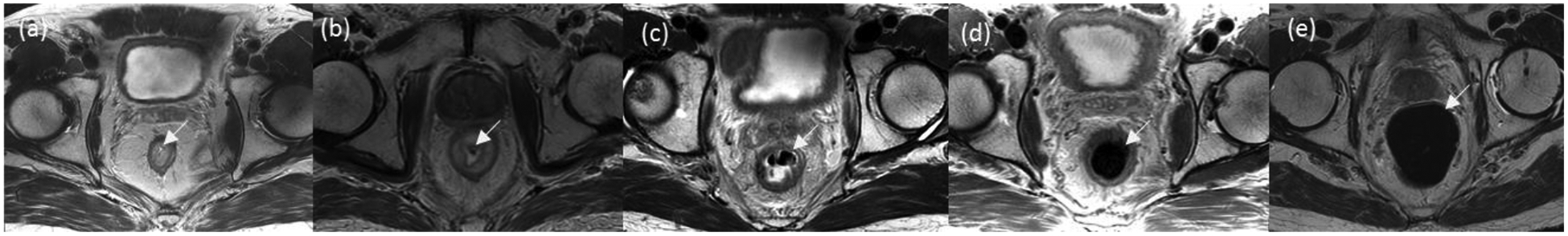
Purpose: To determine whether the administration of a microenema immediately prior to rectal magnetic resonance imaging (MRI) decreases the level of gas-related artifacts on diffusion-weighted imaging (DWI) sequences.
Methods: This retrospective analysis included 492 (183 baseline and 309 post-total neoadjuvant treatment [TNT]) consecutive MRI scans for rectal cancer from January 2019 to January 2020. Scan-related factors were identified including microenema use (yes or no), field of view (FOV) in DWI (b = 800 or b = 1500), and magnet strength (1.5 T or 3 T). Two readers scored DWI studies for gas-related artifacts and T2-weighted sequences for the amount of intraluminal gas on a 5-point scale. Fisher's exact test and the Rao-Scott Chi-squared test were used to examine associations between microenema use and other factors. Generalized estimating equation and multivariable regression models were performed to examine the effect of microenema use in subgroups of scans for each reader. Cohen's κ was used to assess inter-reader agreement.
Results: Gas-related artifact levels decreased in scans with microenema overall (P < 0.001) as well as when scans were stratified by FOV (P ≤ 0.003). For both readers, post-TNT scans with microenema showed lower artifact levels overall (P < 0.014 and P < 0.001) and in post-TNT subgroups of axial DWI scans (P ≤ 0.006 and P < 0.001) and scans acquired with a 3 T magnet (P ≤ 0.001 for both FOV). No evidence of decreased artifact level was found for baseline studies. Decreased gas was seen with microenema use (P < 0.001 for both readers). Inter-reader agreement on artifact-level and gas-level assessments ranged from slight to substantial (κ = 0.273-0.685).
Conclusion: Microenema use prior to rectal MRI reduces gas-related artifacts on DWI, including both large and small FOV sequences and particularly on post-TNT scans performed at 3 T, and offers a viable solution to improve DWI quality.
Keywords: Diffusion-weighted imaging; Magnetic resonance imaging; Microenema; Rectal neoplasms; Susceptibility artifact.
Conflict of interest statement
Figures


References
-
- Howlader N, Noone AM, Krapcho M, Miller D, Brest A, Yu M, Ruhl J, Tatalovich Z, Mariotto A, Lewis DR, Chen HS, Feuer EJ, (eds). CK SEER Cancer Statistics Review, 1975–2017, National Cancer Institute. Bethesda, MD. https://seer.cancer.gov/csr/1975_2017/, based on November 2019 SEER data submission, posted to the SEER web site, April 2020.
-
- Lambregts DM, Vandecaveye V, Barbaro B, Bakers FC, Lambrecht M, Maas M, Haustermans K, Valentini V, Beets GL, Beets-Tan RG (2011) Diffusion-weighted MRI for selection of complete responders after chemoradiation for locally advanced rectal cancer: a multicenter study. Ann Surg Oncol 18 (8):2224–2231. doi:10.1245/s10434-011-1607-5 - DOI - PMC - PubMed
-
- Park MJ, Kim SH, Lee SJ, Jang KM, Rhim H (2011) Locally advanced rectal cancer: added value of diffusion-weighted MR imaging for predicting tumor clearance of the mesorectal fascia after neoadjuvant chemotherapy and radiation therapy. Radiology 260 (3):771–780. doi:10.1148/radiol.11102135 - DOI - PubMed
Publication types
MeSH terms
Grants and funding
LinkOut - more resources
Full Text Sources
