

Docosahexaenoic Acid (DHA) Supplementation Alters Phospholipid Species and Lipid Peroxidation Products in Adult Mouse Brain, Heart, and Plasma
- PMID: 32926329
- PMCID: PMC9555299
- DOI: 10.1007/s12017-020-08616-0
Docosahexaenoic Acid (DHA) Supplementation Alters Phospholipid Species and Lipid Peroxidation Products in Adult Mouse Brain, Heart, and Plasma
Abstract
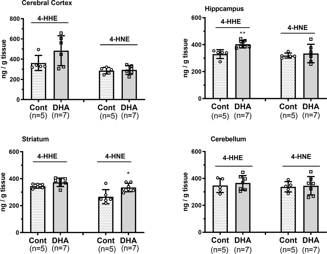
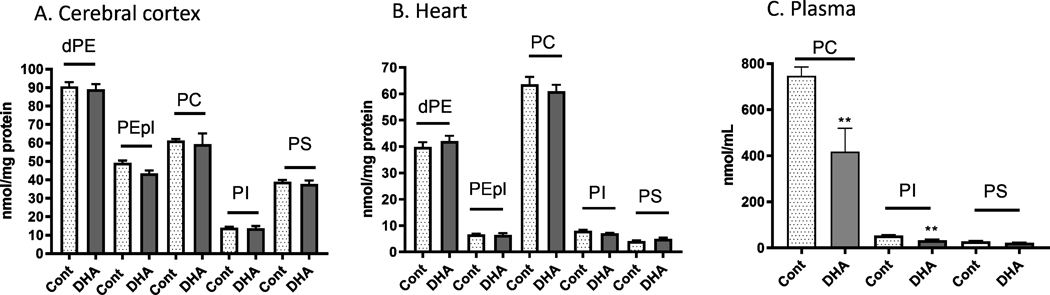
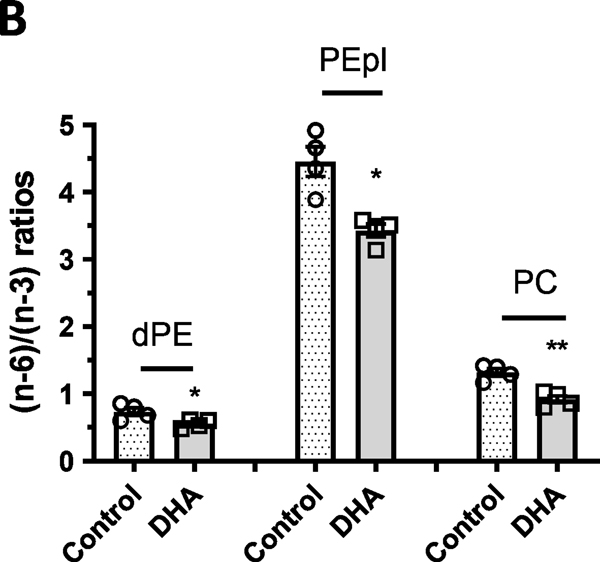
The abundance of docosahexaenoic acid (DHA) in phospholipids in the brain and retina has generated interest to search for its role in mediating neurological functions. Besides the source of many oxylipins with pro-resolving properties, DHA also undergoes peroxidation, producing 4-hydroxyhexenal (4-HHE), although its function remains elusive. Despite wide dietary consumption, whether supplementation of DHA may alter the peroxidation products and their relationship to phospholipid species in brain and other body organs have not been explored sufficiently. In this study, adult mice were administered a control or DHA-enriched diet for 3 weeks, and phospholipid species and peroxidation products were examined in brain, heart, and plasma. Results demonstrated that this dietary regimen increased (n-3) and decreased (n-6) species to different extent in all major phospholipid classes (PC, dPE, PE-pl, PI and PS) examined. Besides changes in phospholipid species, DHA-enriched diet also showed substantial increases in 4-HHE in brain, heart, and plasma. Among different brain regions, the hippocampus responded to the DHA-enriched diet showing significant increase in 4-HHE. Considering the pro- and anti-inflammatory pathways mediated by the (n-6) and (n-3) polyunsaturated fatty acids, unveiling the ability for DHA-enriched diet to alter phospholipid species and lipid peroxidation products in the brain and in different body organs may be an important step forward towards understanding the mechanism(s) for this (n-3) fatty acid on health and diseases.
Keywords: Docosahexaenoic acid; Heart; Hippocampus; Lipid peroxidation; Lipidomics; Plasma.
Conflict of interest statement
DECLARATIONS
Figures


Similar articles
-
Unveiling anti-oxidative and anti-inflammatory effects of docosahexaenoic acid and its lipid peroxidation product on lipopolysaccharide-stimulated BV-2 microglial cells.J Neuroinflammation. 2018 Jul 9;15(1):202. doi: 10.1186/s12974-018-1232-3. J Neuroinflammation. 2018. PMID: 29986724 Free PMC article.
-
Dietary DHA supplementation causes selective changes in phospholipids from different brain regions in both wild type mice and the Tg2576 mouse model of Alzheimer's disease.Biochim Biophys Acta. 2016 Jun;1861(6):524-37. doi: 10.1016/j.bbalip.2016.03.005. Epub 2016 Mar 9. Biochim Biophys Acta. 2016. PMID: 26968097 Free PMC article.
-
Maternal Dietary Docosahexaenoic Acid Alters Lipid Peroxidation Products and (n-3)/(n-6) Fatty Acid Balance in Offspring Mice.Metabolites. 2019 Mar 1;9(3):40. doi: 10.3390/metabo9030040. Metabolites. 2019. PMID: 30832208 Free PMC article.
-
Docosahexaenoic acid (DHA): An essential nutrient and a nutraceutical for brain health and diseases.Prostaglandins Leukot Essent Fatty Acids. 2018 Sep;136:3-13. doi: 10.1016/j.plefa.2017.03.006. Epub 2017 Mar 10. Prostaglandins Leukot Essent Fatty Acids. 2018. PMID: 28314621 Free PMC article. Review.
-
Yin-Yang Mechanisms Regulating Lipid Peroxidation of Docosahexaenoic Acid and Arachidonic Acid in the Central Nervous System.Front Neurol. 2019 Jun 18;10:642. doi: 10.3389/fneur.2019.00642. eCollection 2019. Front Neurol. 2019. PMID: 31275232 Free PMC article. Review.
Cited by
-
Encapsulation of Docosahexaenoic Acid Oil Substantially Improves the Oxylipin Profile of Rat Tissues.Front Nutr. 2022 Jan 13;8:812119. doi: 10.3389/fnut.2021.812119. eCollection 2021. Front Nutr. 2022. PMID: 35118110 Free PMC article.
-
Lipidomic analysis of brain and hippocampus from mice fed with high-fat diet and treated with fecal microbiota transplantation.Nutr Metab (Lond). 2023 Feb 15;20(1):12. doi: 10.1186/s12986-023-00730-7. Nutr Metab (Lond). 2023. PMID: 36793054 Free PMC article.
-
Dynamic Role of Phospholipases A2 in Health and Diseases in the Central Nervous System.Cells. 2021 Oct 30;10(11):2963. doi: 10.3390/cells10112963. Cells. 2021. PMID: 34831185 Free PMC article. Review.
-
Effect of Maternal Dietary DHA and Prenatal Stress Mouse Model on Autistic-like Behaviors, Lipid Peroxidation Activity, and GABA Expression in Offspring Pups.Int J Mol Sci. 2025 Jul 14;26(14):6730. doi: 10.3390/ijms26146730. Int J Mol Sci. 2025. PMID: 40724979 Free PMC article.
References
-
- Bascoul-Colombo C, Guschina IA, Maskrey BH, Good M, O’Donnell VB, & Harwood JL (2016). Dietary DHA supplementation causes selective changes in phospholipids from different brain regions in both wild type mice and the Tg2576 mouse model of Alzheimer’s disease. Biochim Biophys Acta, 1861(6), 524–537. doi:10.1016/j.bbalip.2016.03.005 - DOI - PMC - PubMed
Publication types
MeSH terms
Substances
Grants and funding
LinkOut - more resources
Full Text Sources
Medical
Research Materials
Miscellaneous
