Meta-Analysis
. 2021 Jan;56(1):252-263.
doi: 10.1002/ppul.25070. Epub 2020 Nov 2.

Pediatric lung imaging features of COVID-19: A systematic review and meta-analysis

Affiliations
Meta-Analysis

Pediatric lung imaging features of COVID-19: A systematic review and meta-analysis

Gustavo Nino et al. Pediatr Pulmonol. 2021 Jan.

Abstract

Rationale: Pediatric COVID-19 studies have been mostly restricted to case reports and small case series, which have prevented the identification of specific pediatric lung disease patterns in COVID-19. The overarching goal of this systematic review and meta-analysis is to provide the first comprehensive summary of the findings of published studies thus far describing COVID-19 lung imaging data in the pediatric population.

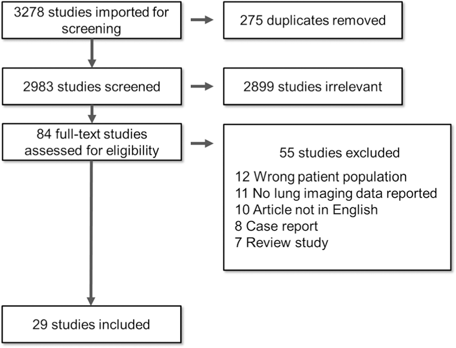
Methods: A systematic literature search of PubMed was performed to identify studies assessing lung-imaging features of COVID-19 pediatric patients (0-18 years). A single-arm meta-analysis was conducted to obtain the pooled prevalence and 95% confidence interval (95% CI).

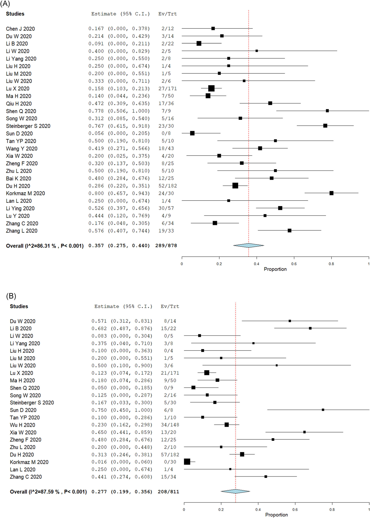
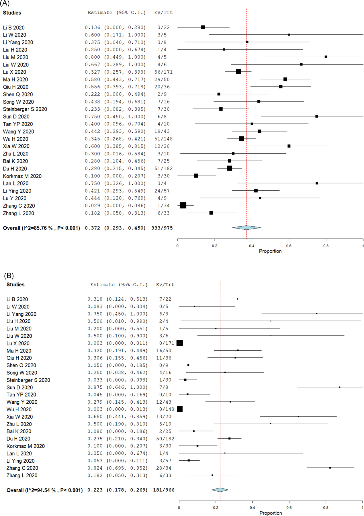
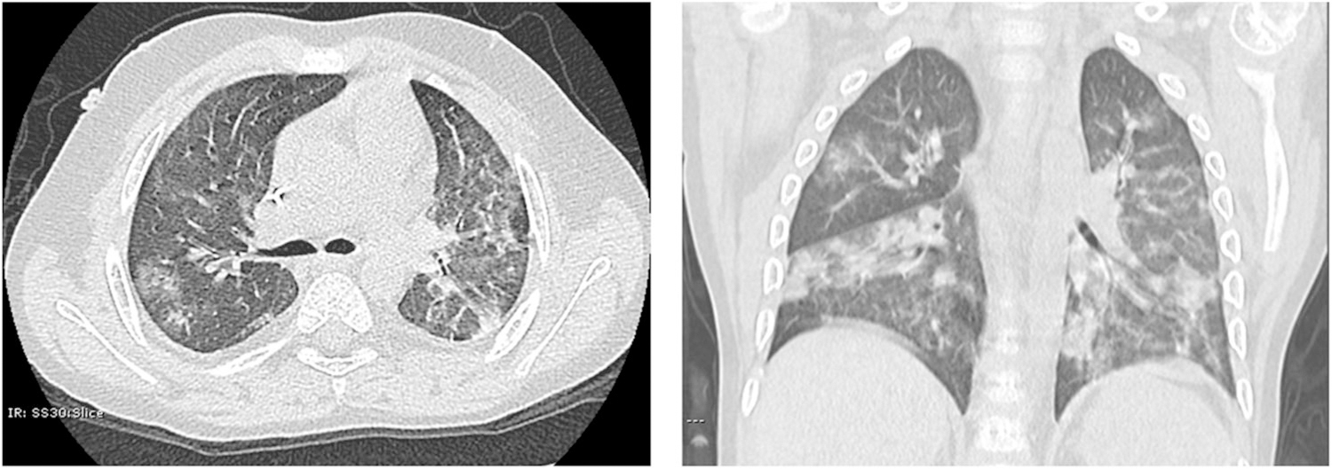
Results: A total of 29 articles (n = 1026 children) based on chest computerized tomography (CT) images were included. The main results of this comprehensive analysis are as follows: (1) Over a third of pediatric patients with COVID-19 (35.7%, 95% CI: 27.5%-44%) had normal chest CT scans and only 27.7% (95% CI: 19.9%-35.6%) had bilateral lesions. (2) The most typical pediatric chest CT findings of COVID-19 were ground-glass opacities (GGO) (37.2%, 95% CI: 29.3%-45%) and the presence of consolidations or pneumonic infiltrates (22.3%, 95% CI: 17.8%-26.9%). (3) The lung imaging findings in children with COVID-19 were overall less frequent and less severe than in adult patients. (4) Typical lung imaging features of viral respiratory infections in the pediatric population such as increased perihilar markings and hyperinflation were not reported in children with COVID-19.

Conclusion: Chest CT manifestations in children with COVID-19 could potentially be used for early identification and prompt intervention in the pediatric population.

Keywords: SARS-CoV-2; lung CT scan; meta-analysis; pediatric COVID-19.


Conflict of interest statement

CONFLICT OF INTERESTS

The authors declare that there are no conflict of interests.

Figures

FIGURE 1
FIGURE 1
Diagram of document retrieval
FIGURE 2
FIGURE 2
Lung imaging results in pediatric COVID-19 cases. Forest plots for single-arm meta-analysis of the pediatric COVID-19 studies reporting (A) normal lung imaging; or (B) bilateral compromise. Data presented as the 95% confidence interval (CI) of the proportion of subjects with normal findings or bilateral compromise in each study. COVID-19, coronavirus disease 2019 [Color figure can be viewed at wileyonlinelibrary.com]
FIGURE 3
FIGURE 3
Top lung imaging abnormalities in pediatric COVID-19 cases. Forest plots for single-arm meta-analysis of the pediatric COVID-19 studies reporting (A) ground-glass opacifications (GGO); or (B) consolidations or pneumonic infiltrates. Data presented as the 95% confidence interval (CI) of the proportion of subjects with these lung abnormalities in each study. COVID-19, coronavirus disease 2019 [Color figure can be viewed at wileyonlinelibrary.com]
FIGURE 4
FIGURE 4
Typical radiological appearance of the most common abnormalities identified in pediatric COVID-19 cases. Chest CT of a 5-year-old male with PCR-confirmed SARS-CoV-2 infection. Images show multifocal scattered GGO and patchy consolidations with a peripheral distribution. There is no hyperinflation, increased perihilar markings, pleural thickening, pleural effusion, or bronchiectasis present. COVID-19, coronavirus disease 2019; CT, computerized tomography; GGO, ground-glass opacity; PCR, polymerase chain reaction; SARS-CoV-2, severe acute respiratory syndrome coronavirus 2

References

    1. Chen R, Liang W, Medical Treatment Expert Group for COVID-19, et al. Risk factors of fatal outcome in hospitalized subjects with coronavirus disease 2019 from a nationwide analysis in China. Chest. 2020;158(1):97–105. 10.1016/j.chest.2020.04.010 - DOI - PMC - PubMed
    1. Gudbjartsson DF, Helgason A, Jonsson H, et al. Spread of SARS-CoV-2 in the Icelandic population. N Engl J Med. 2020;382: 2302–2315. 10.1056/NEJMoa2006100 - DOI - PMC - PubMed
    1. CDC COVID-19 Response Team. Coronavirus disease 2019 in Children—United States, February 12–April 2, 2020. MMWR Morb Mortal Wkly Rep. 2020;69(14):422–426. 10.15585/mmwr.mm6914e4 - DOI - PMC - PubMed
    1. Tagarro A, Epalza C, Santos M, et al. Screening and severity of coronavirus disease 2019 (COVID-19) in children in Madrid, Spain. JAMA Pediatr. 2020;April 8:e201346. 10.1001/jamapediatrics.2020.1346 [Epub ahead of print]. PubMed PMID: 32267485; PubMed Central PMCID: PMC7142799. - DOI - PMC - PubMed
    1. Morand A, Fabre A, Minodier P, et al. COVID-19 virus and children: What do we know? Arch Pediatr. 2020;27(3):117–118. 10.1016/j.arcped.2020.03.001 - DOI - PMC - PubMed

Publication types