

Atypical Cause of Sepsis from Bilateral Iliopsoas Abscesses Seeded from Self-mutilation: A Case Report
- PMID: 32926705
- PMCID: PMC7434279
- DOI: 10.5811/cpcem.2020.5.47020
Atypical Cause of Sepsis from Bilateral Iliopsoas Abscesses Seeded from Self-mutilation: A Case Report
Abstract
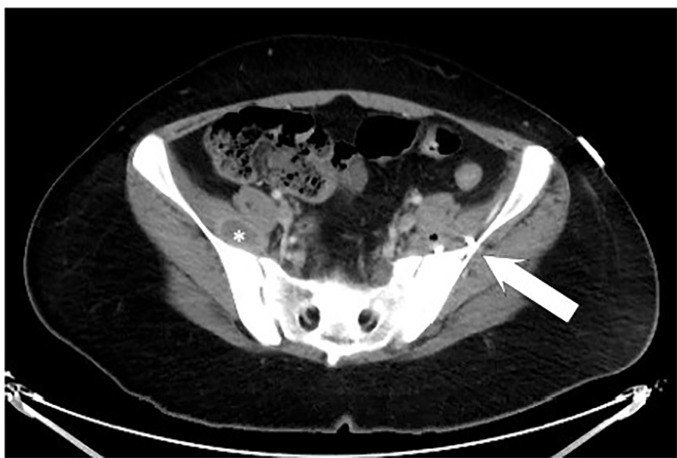
Introduction: An iliopsoas abscess (IPA) is an abscess located adjacent to the iliopsoas and iliacus muscles. Although rare, their variable clinical presentations often lead to a delay in diagnosis.
Case report: We present a case of sepsis secondary to multiple IPAs that was missed despite multiple healthcare encounters. The patient had no classical risk factors for an IPA, and the abscesses were found to be seeded via hematogenous spread from self-inflicted cutting.
Conclusion: This case illustrates the importance of obtaining a complete history, including psychiatric screen, and performing a thorough examination when evaluating patients with low back pain to rule out overlooked sources of bacteremia.
Conflict of interest statement
Figures


Similar articles
-
Primary Iliopsoas Abscess and Drug-Induced Liver Injury in the Emergency Department: A Case Report.Diseases. 2024 Dec 12;12(12):326. doi: 10.3390/diseases12120326. Diseases. 2024. PMID: 39727656 Free PMC article.
-
A Case Report of Iliopsoas Abscess Secondary to Small Bowel Fistula.Cureus. 2023 Feb 7;15(2):e34749. doi: 10.7759/cureus.34749. eCollection 2023 Feb. Cureus. 2023. PMID: 36909091 Free PMC article.
-
Epidural phlegmon and iliopsoas abscess caused by Salmonella enterica bacteremia: A case report.Int J Surg Case Rep. 2022 Jul;96:107287. doi: 10.1016/j.ijscr.2022.107287. Epub 2022 Jun 7. Int J Surg Case Rep. 2022. PMID: 35696819 Free PMC article.
-
Iliopsoas abscesses: diagnostic, aetiologic and therapeutic approach in five patients with a literature review.Scand J Gastroenterol. 2009;44(5):594-9. doi: 10.1080/00365520902745054. Scand J Gastroenterol. 2009. PMID: 19225988 Review.
-
Lemierre's disease: a case with bilateral iliopsoas abscesses and a literature review.World J Emerg Surg. 2014 May 15;9:38. doi: 10.1186/1749-7922-9-38. eCollection 2014. World J Emerg Surg. 2014. PMID: 24904685 Free PMC article. Review.
References
-
- Ricci MA, Rose FB, Meyer KK. Pyogenic psoas abscess: worldwide variations in etiology. World J Surg. 1986;10(5):834–42. - PubMed
-
- Bartolo DC, Ebbs SR, Cooper MJ. Psoas abscess in Bristol: a 10-year review. Int J Colorectal Dis. 1987;2(2):72–6. - PubMed
-
- Garner JP, Meiring PD, Ravi K, et al. Psoas abscess – not as rare as we think? Colorectal Dis. 2007;9(3):269–74. - PubMed
-
- Dinç H, Onder C, Turhan AU, et al. Percutaneous catheter drainage of tuberculous and nontuberculous psoas abscesses. Eur J Radiol. 1996;23(2):130–4. - PubMed
-
- Agrawal SN, Dwivedi AJ, Khan M. Primary psoas abscess. Dig Dis Sci. 2002;47(9):2103–5. - PubMed