Observational Study
. 2021 Jun;5(6):580-592.
doi: 10.1016/j.oret.2020.09.004. Epub 2020 Sep 11.

Macular OCT Characteristics at 36 Weeks' Postmenstrual Age in Infants Examined for Retinopathy of Prematurity

Affiliations
Observational Study

Macular OCT Characteristics at 36 Weeks' Postmenstrual Age in Infants Examined for Retinopathy of Prematurity

Shwetha Mangalesh et al. Ophthalmol Retina. 2021 Jun.

Abstract

Purpose: To report our ability to capture,-grade reliably, and analyze bedside macular OCT images from preterm infants and relate OCT findings to biological factors and retinopathy of prematurity (ROP) status at a single time window in the Study of Eye Imaging in Preterm Infants (BabySTEPS).

Design: Prospective, observational study.

Participants: Preterm infants eligible for ROP screening with parental consent for research and a 36 ± 1 weeks' postmenstrual age (PMA) visit.

Methods: We imaged both eyes of preterm infants with an investigational noncontact, handheld swept-source (SS) OCT at the time of clinical ROP examinations. Macular OCT features and layer thicknesses for untreated eyes of infants at 36 ± 1 weeks' PMA were compared with demographic data and clinical ROP examination performed by experts. Statistical analyses accounted for the use of both eyes of infants.

Main outcome measures: Macular OCT features and layer thicknesses, gender, race or ethnicity, gestational age, birth weight, ROP stage, and plus disease.

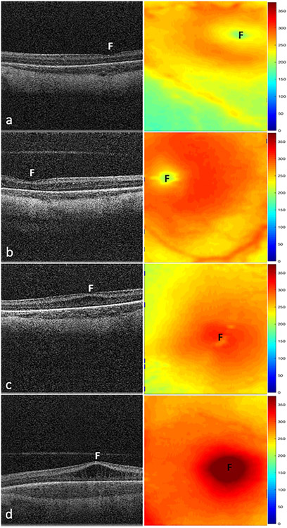
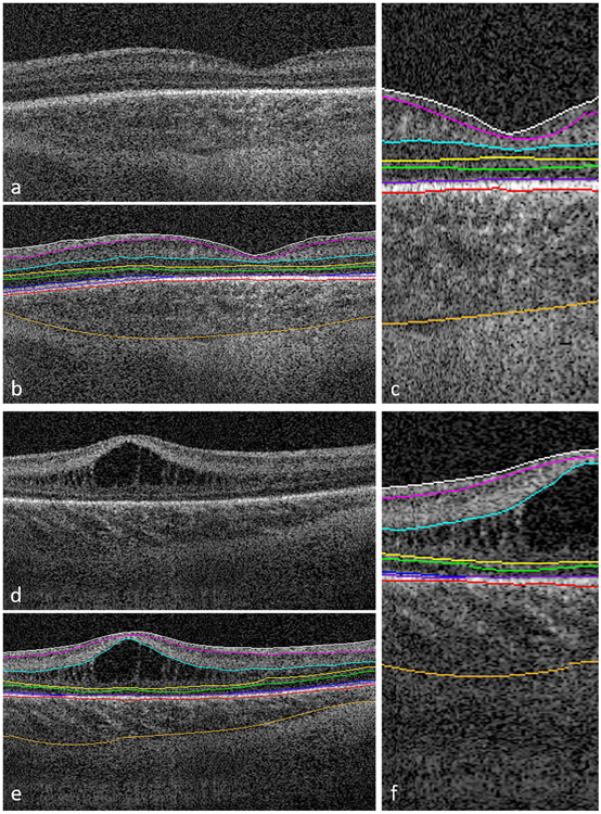
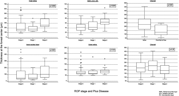
Results: We captured macular OCT from 169 eyes (1 eye excluded because of prior ROP treatment) at 36 ± 1 weeks' PMA. The quality of OCT volumes was excellent in 33 eyes (19%), acceptable in 112 eyes (67%), poor in 24 eyes (14%), and unusable in 0 eyes (0%). Macular edema was present in 60% of eyes and was bilateral in 82% of infants with edema. At the fovea, retinal and inner nuclear layer thickness increased with edema severity: 183 ± 36 μm and 51 ± 27 μm in mild (16% of eyes), 308 ± 57 μm and 163 ± 53 μm in moderate (25%), and 460 ± 76 μm and 280 ± 83 μm in severe edema (12%), respectively. With an increase in ROP stage from 0 to 2, the mean ± standard deviation retinal thickness at the fovea increased from 227± 124 μm to 297 ± 99 μm (P < 0.001). The choroid was thinner, 155 ± 72 μm, with preplus or plus disease versus without, 236 ± 79 μm (P = 0.04), whereas retinal thickness did not vary.

Conclusions: We demonstrated the reliability of methods and the prevalence of OCT findings in preterm infants enrolled in BabySTEPS at a single time point of 36 ± 1 weeks' PMA. Variations in layer thicknesses in infants at this time point may reflect abnormalities resulting from delay in foveal development that may be impacted by macular edema, ROP, or both.

Keywords: BabySTEPS; OCT; Preterm infants; Retinopathy of prematurity; swept-source OCT.


Conflict of interest statement

Financial Disclosures: Dr. Toth receives royalties through her university from Alcon and Hemosonics. Dr. Toth has unlicensed and pending patents related to the investigational device and imaging in this study. Drs. Viehland, Izatt and Toth have a patent application pending on the novel handheld probe (investigational handheld UC3 system) described in this manuscript. Dr. Izatt and Duke University have licensed technology to and have a financial interest (including royalty/milestone payments) in Bioptigen/Leica Microsystems Inc., which manufactures handheld and intrasurgical OCT systems. Dr. Vajzovic has received research funding from Heidelberg Engineering Inc., Orbit Biomedical Inc., Novartis and Second Sight Inc. and has served as a consultant to AERI, Alcon, Alimera Sciences, Allergan, Baush and Lomb, DORC, Genentech, Guidepoint, Janssen Pharmaceutical, Orbit Biomedical and Second Sight. No other authors have related conflict of interest. No authors have a proprietary interest in the current study.

Figures

Figure 1.
Figure 1.
Representative swept-source OCT B-scans (left column) and retinal thickness maps (right column) of macular edema severity in stage 2 ROP. A, Infant with no edema with a retinal thickness of 186 μm and inner nuclear layer (INL) thickness of 30 μm at the fovea (F). B, Infant with mild edema showing little to no deformation of the foveal contour with a retinal thickness of 212 μm and INL thickness of 52 μm. C, Infant with moderate edema showing flattening or slight upward bulging of the fovea and retina and INL thicknesses measuring 294 μm and 151 μm, respectively. D, Infant with severe edema showing severe upward bulging of the foveal contour with retinal and INL thicknesses of 433 μm and 286 μm, respectively. Mean retinal thickness at the fovea increased from 160 μm (standard deviation, 43 μm) without macular edema to 460 μm (standard deviation, 76 μm) with severe macular edema (P < 0.001).
Figure 2.
Figure 2.
Representative foveal B-scans from macular volumes from swept-source OCT systems in infants with (A, B, C) no macular edema and (D, E, F) with macular edema. B, C, E, F, Duke OCT Retinal Analysis Program Marking Code Baby version 2.0 semiautomated segmentation at the internal limiting membrane (white), outer borders of the nerve fiber layer (magenta), inner plexiform layer (aqua), inner nuclear layer (yellow), outer plexiform layer (green), ellipsoid zone (blue; not visualized in (C) and tapering at the foveal margin in (F)), retinal pigment epithelium (inner, purple; outer, pink), and choroid (orange).
Figure 3:
Figure 3:
Box-whisker plots showing retinal layer and choroidal thicknesses across retinopathy of prematurity stages and choroidal thickness association with pre-plus and plus disease at the fovea center. Thicker total retina, retinal nerve fiber layer (RNFL)+ganglion cell layer (GCL)+inner plexiform layer (IPL), and inner nuclear layer were associated with a higher ROP stage, and the choroid was thinner in infants with preplus and plus disease.

References

    1. Patel CK. Optical coherence tomography in the management of acute retinopathy of prematurity. Am J Ophthalmol 2006;141(3):582–4. - PubMed
    1. Vinekar A, Sivakumar M, Shetty R, et al. A novel technique using spectral-domain optical coherence tomography (Spectralis, SD-OCT+HRA) to image supine non-anaesthetized infants: utility demonstrated in aggressive posterior retinopathy of prematurity. Eye (Lond) 2010;24(2):379–82. - PubMed
    1. Maldonado RS, Izatt JA, Sarin N, et al. Optimizing hand-held spectral domain optical coherence tomography imaging for neonates, infants, and children. Invest Ophthalmol Vis Sci 2010;51(5):2678–85. - PMC - PubMed
    1. Lee AC, Maldonado RS, Sarin N, et al. Macular features from spectral-domain optical coherence tomography as an adjunct to indirect ophthalmoscopy in retinopathy of prematurity. Retina 2011;31(8):1470–82. - PMC - PubMed
    1. Chavala SH, Farsiu S, Maldonado R, et al. Insights into advanced retinopathy of prematurity using handheld spectral domain optical coherence tomography imaging. Ophthalmology 2009;116(12):2448–56. - PMC - PubMed

Publication types

LinkOut - more resources